WO2003106495A2 - MONOCLONAL ANTIBODY hPAM4 - Google Patents
MONOCLONAL ANTIBODY hPAM4 Download PDFInfo
- Publication number
- WO2003106495A2 WO2003106495A2 PCT/GB2003/002593 GB0302593W WO03106495A2 WO 2003106495 A2 WO2003106495 A2 WO 2003106495A2 GB 0302593 W GB0302593 W GB 0302593W WO 03106495 A2 WO03106495 A2 WO 03106495A2
- Authority
- WO
- WIPO (PCT)
- Prior art keywords
- antibody
- fragment
- pam4
- diagnostic
- agent
- Prior art date
Links
- 0 C*NC=*Nc1ccc(CC2*(CC(O)=O)CCN(CC(O)=O)CCN(CC(O)=O)C2)cc1 Chemical compound C*NC=*Nc1ccc(CC2*(CC(O)=O)CCN(CC(O)=O)CCN(CC(O)=O)C2)cc1 0.000 description 1
Classifications
-
- A—HUMAN NECESSITIES
- A61—MEDICAL OR VETERINARY SCIENCE; HYGIENE
- A61K—PREPARATIONS FOR MEDICAL, DENTAL OR TOILETRY PURPOSES
- A61K49/00—Preparations for testing in vivo
- A61K49/0004—Screening or testing of compounds for diagnosis of disorders, assessment of conditions, e.g. renal clearance, gastric emptying, testing for diabetes, allergy, rheuma, pancreas functions
- A61K49/0008—Screening agents using (non-human) animal models or transgenic animal models or chimeric hosts, e.g. Alzheimer disease animal model, transgenic model for heart failure
-
- C—CHEMISTRY; METALLURGY
- C07—ORGANIC CHEMISTRY
- C07K—PEPTIDES
- C07K16/00—Immunoglobulins [IGs], e.g. monoclonal or polyclonal antibodies
- C07K16/18—Immunoglobulins [IGs], e.g. monoclonal or polyclonal antibodies against material from animals or humans
-
- A—HUMAN NECESSITIES
- A61—MEDICAL OR VETERINARY SCIENCE; HYGIENE
- A61K—PREPARATIONS FOR MEDICAL, DENTAL OR TOILETRY PURPOSES
- A61K47/00—Medicinal preparations characterised by the non-active ingredients used, e.g. carriers or inert additives; Targeting or modifying agents chemically bound to the active ingredient
- A61K47/50—Medicinal preparations characterised by the non-active ingredients used, e.g. carriers or inert additives; Targeting or modifying agents chemically bound to the active ingredient the non-active ingredient being chemically bound to the active ingredient, e.g. polymer-drug conjugates
- A61K47/51—Medicinal preparations characterised by the non-active ingredients used, e.g. carriers or inert additives; Targeting or modifying agents chemically bound to the active ingredient the non-active ingredient being chemically bound to the active ingredient, e.g. polymer-drug conjugates the non-active ingredient being a modifying agent
- A61K47/68—Medicinal preparations characterised by the non-active ingredients used, e.g. carriers or inert additives; Targeting or modifying agents chemically bound to the active ingredient the non-active ingredient being chemically bound to the active ingredient, e.g. polymer-drug conjugates the non-active ingredient being a modifying agent the modifying agent being an antibody, an immunoglobulin or a fragment thereof, e.g. an Fc-fragment
-
- A—HUMAN NECESSITIES
- A61—MEDICAL OR VETERINARY SCIENCE; HYGIENE
- A61K—PREPARATIONS FOR MEDICAL, DENTAL OR TOILETRY PURPOSES
- A61K51/00—Preparations containing radioactive substances for use in therapy or testing in vivo
- A61K51/02—Preparations containing radioactive substances for use in therapy or testing in vivo characterised by the carrier, i.e. characterised by the agent or material covalently linked or complexing the radioactive nucleus
- A61K51/04—Organic compounds
- A61K51/08—Peptides, e.g. proteins, carriers being peptides, polyamino acids, proteins
- A61K51/10—Antibodies or immunoglobulins; Fragments thereof, the carrier being an antibody, an immunoglobulin or a fragment thereof, e.g. a camelised human single domain antibody or the Fc fragment of an antibody
- A61K51/1045—Antibodies or immunoglobulins; Fragments thereof, the carrier being an antibody, an immunoglobulin or a fragment thereof, e.g. a camelised human single domain antibody or the Fc fragment of an antibody against animal or human tumor cells or tumor cell determinants
- A61K51/1057—Antibodies or immunoglobulins; Fragments thereof, the carrier being an antibody, an immunoglobulin or a fragment thereof, e.g. a camelised human single domain antibody or the Fc fragment of an antibody against animal or human tumor cells or tumor cell determinants the tumor cell being from liver or pancreas
-
- A—HUMAN NECESSITIES
- A61—MEDICAL OR VETERINARY SCIENCE; HYGIENE
- A61P—SPECIFIC THERAPEUTIC ACTIVITY OF CHEMICAL COMPOUNDS OR MEDICINAL PREPARATIONS
- A61P35/00—Antineoplastic agents
-
- A—HUMAN NECESSITIES
- A61—MEDICAL OR VETERINARY SCIENCE; HYGIENE
- A61P—SPECIFIC THERAPEUTIC ACTIVITY OF CHEMICAL COMPOUNDS OR MEDICINAL PREPARATIONS
- A61P37/00—Drugs for immunological or allergic disorders
-
- A—HUMAN NECESSITIES
- A61—MEDICAL OR VETERINARY SCIENCE; HYGIENE
- A61P—SPECIFIC THERAPEUTIC ACTIVITY OF CHEMICAL COMPOUNDS OR MEDICINAL PREPARATIONS
- A61P37/00—Drugs for immunological or allergic disorders
- A61P37/02—Immunomodulators
-
- A—HUMAN NECESSITIES
- A61—MEDICAL OR VETERINARY SCIENCE; HYGIENE
- A61P—SPECIFIC THERAPEUTIC ACTIVITY OF CHEMICAL COMPOUNDS OR MEDICINAL PREPARATIONS
- A61P43/00—Drugs for specific purposes, not provided for in groups A61P1/00-A61P41/00
-
- C—CHEMISTRY; METALLURGY
- C07—ORGANIC CHEMISTRY
- C07K—PEPTIDES
- C07K14/00—Peptides having more than 20 amino acids; Gastrins; Somatostatins; Melanotropins; Derivatives thereof
- C07K14/435—Peptides having more than 20 amino acids; Gastrins; Somatostatins; Melanotropins; Derivatives thereof from animals; from humans
- C07K14/46—Peptides having more than 20 amino acids; Gastrins; Somatostatins; Melanotropins; Derivatives thereof from animals; from humans from vertebrates
- C07K14/47—Peptides having more than 20 amino acids; Gastrins; Somatostatins; Melanotropins; Derivatives thereof from animals; from humans from vertebrates from mammals
- C07K14/4701—Peptides having more than 20 amino acids; Gastrins; Somatostatins; Melanotropins; Derivatives thereof from animals; from humans from vertebrates from mammals not used
- C07K14/4748—Tumour specific antigens; Tumour rejection antigen precursors [TRAP], e.g. MAGE
-
- C—CHEMISTRY; METALLURGY
- C07—ORGANIC CHEMISTRY
- C07K—PEPTIDES
- C07K16/00—Immunoglobulins [IGs], e.g. monoclonal or polyclonal antibodies
- C07K16/18—Immunoglobulins [IGs], e.g. monoclonal or polyclonal antibodies against material from animals or humans
- C07K16/28—Immunoglobulins [IGs], e.g. monoclonal or polyclonal antibodies against material from animals or humans against receptors, cell surface antigens or cell surface determinants
- C07K16/2896—Immunoglobulins [IGs], e.g. monoclonal or polyclonal antibodies against material from animals or humans against receptors, cell surface antigens or cell surface determinants against molecules with a "CD"-designation, not provided for elsewhere
-
- C—CHEMISTRY; METALLURGY
- C07—ORGANIC CHEMISTRY
- C07K—PEPTIDES
- C07K16/00—Immunoglobulins [IGs], e.g. monoclonal or polyclonal antibodies
- C07K16/18—Immunoglobulins [IGs], e.g. monoclonal or polyclonal antibodies against material from animals or humans
- C07K16/28—Immunoglobulins [IGs], e.g. monoclonal or polyclonal antibodies against material from animals or humans against receptors, cell surface antigens or cell surface determinants
- C07K16/30—Immunoglobulins [IGs], e.g. monoclonal or polyclonal antibodies against material from animals or humans against receptors, cell surface antigens or cell surface determinants from tumour cells
-
- A—HUMAN NECESSITIES
- A61—MEDICAL OR VETERINARY SCIENCE; HYGIENE
- A61K—PREPARATIONS FOR MEDICAL, DENTAL OR TOILETRY PURPOSES
- A61K39/00—Medicinal preparations containing antigens or antibodies
- A61K2039/505—Medicinal preparations containing antigens or antibodies comprising antibodies
-
- C—CHEMISTRY; METALLURGY
- C07—ORGANIC CHEMISTRY
- C07K—PEPTIDES
- C07K2317/00—Immunoglobulins specific features
- C07K2317/20—Immunoglobulins specific features characterized by taxonomic origin
- C07K2317/24—Immunoglobulins specific features characterized by taxonomic origin containing regions, domains or residues from different species, e.g. chimeric, humanized or veneered
Definitions
- This invention relates to monovalent and multivalent, monospecific antibodies and to multivalent, multispecific antibodies.
- the present invention relates to a MUCI antigen specific antibody designated PAM4.
- the invention further relates to humanized and human PAM4 antibodies and fragments thereof, and the use of such antibodies and fragments thereof in diagnosis and therapy.
- the antibodies of the present invention have one or more identical binding sites, wherein each binding site has an affinity toward a target antigen or an epitope on a target antigen. In another embodiment, the antibodies of the present invention have two or more binding sites which have an affinity toward the same or different epitopes on a target antigen or the same or different target antigens, or at least one binding site has an affinity toward a target antigen and at least one binding site has an affinity toward a hapten.
- the present invention also describes recombinant vectors useful for expressing the antibodies described herein in a host.
- pancreas produces insulin to assist the body in converting glucose to energy and enzymes to assist the body in digesting food.
- Pancreatic cancer is a malignant growth of the pancreas that mainly occurs in the cells of the pancreatic ducts. This disease is the ninth most common form of cancer, yet it is the fourth and fifth leading cause of cancer deaths in men and women, respectively. Cancer of the pancreas is almost always fatal, with a five-year survival rate that is less than 3%.
- pancreatic cancer The most common symptoms of pancreatic cancer include jaundice, abdominal pain, and weight loss, which, together with other presenting factors, are nonspecific in nature. Thus, diagnosing pancreatic cancer at an early stage of tumor growth is often difficult and requires considerable suspicion and extensive diagnostic work-up, often times including exploratory surgery. Endoscopic ultrasonography and computed tomography are the best noninvasive means available today for diagnosis of pancreatic cancer. However, reliable detection of small tumors, as well as differentiation of pancreatic cancer from focal pancreatitis, is troublesome. Unfortunately, the vast majority of patients are presently diagnosed at a late stage when the tumor has already extended outside of the capsule to invade surrounding organs and/or has metastasized extensively. Gold et at., Crit. Rev. Oncology/Hematology, 39:147-54 (2001). Late detection of the disease is common, and "early" pancreatic cancer diagnosis is rare in the clinical setting.
- pancreatic cancer Early detection and diagnosis of pancreatic cancer, as well as appropriate staging of the disease, would provide an increased survival advantage.
- a number of laboratories are proceeding on the development of a diagnostic procedure based upon the release of a tumor- associated marker into the bloodstream as well as detection of the marker substance within biopsy specimens.
- the best tumor associated marker for pancreatic cancer has been the immunoassay for CA19.9. Elevated levels of this sialylated Le a epitope structure were found in 70% of pancreatic cancer patients but were not found in any of the focal pancreatitis specimens examined. However, CA19.9 levels were found to be elevated in a number of other malignant and benign conditions, so that currently the assay cannot be used for diagnosis.
- MAbs monoclonal antibodies
- DUPAN2, SPAN1, B72.3, Ia3, and various anti-CEA antibodies include but are not limited to DUPAN2, SPAN1, B72.3, Ia3, and various anti-CEA antibodies.
- Man-made antibodies in particular MAbs and engineered antibodies or antibody fragments, have been tested widely and shown to be of value in detection and treatment of pancreatic cancer, as well as other various human disorders, including cancers, autoimmune diseases, infectious diseases, inflammatory diseases, and cardiovascular diseases [Filpula and McGuire, Exp. Opin. Ther. Patents (1999) 9: 231-245].
- the clinical utility of an antibody or an antibody-derived agent is primarily dependent on its ability to bind to a specific targeted antigen associated with a specific disorder.
- a diagnostic or therapeutic agent such as isotopes, drugs, toxins, cytokines, hormones, hormone antagonists, enzymes, enzyme inhibitors, oligonucleotides, growth factors, oligonucleotides, radionuclides, an angiogenesis inhibitor, or metals, to a target location during the detection and treatment phases of a human disorder, particularly if the diagnostic or therapeutic agent is toxic to normal tissue in the body.
- adiolabeled antibodies have been used with some success in numerous malignancies, including ovarian cancer, colon cancer and lymphoma. This technology may also prove useful for pancreatic cancer.
- anti-CEA antibodies and B72.3 little clinical information exists.
- the important parameters in the detection and treatment techniques are the amount of the injected dose specifically localized at the site(s) where target cells are present and the uptake ratio, i.e. the ratio of the concentration of specifically bound antibody to that of the radioactivity present in surrounding normal tissues.
- the uptake ratio i.e. the ratio of the concentration of specifically bound antibody to that of the radioactivity present in surrounding normal tissues.
- an antibody When an antibody is injected into the blood stream, it passes through a number of compartments as it is metabolized and excreted. The antibody must be able to locate and bind to the target cell antigen while passing through the rest of the body.
- Factors that control antigen targeting include location, size, antigen density, antigen accessibility, cellular composition of pathologic tissue, and the pharmacokinetics of the targeting antibodies.
- Target antigens both in tumor and other tissues
- bone marrow toxicity resulting from the slow blood-clearance of the radiolabeled antibodies.
- the amount of targeting antibodies accreted by the targeted tumor cells is influenced by the vascularization and barriers to antibody penetration of tumors, as well as intratumoral pressure.
- Non-specific uptake by non-target organs such as the liver, kidneys or bone-marrow is another potential limitation of the technique, especially for radioimmunotherapy, where irradiation of the bone marrow often causes the dose-limiting toxicity.
- direct targeting is a technique designed to target specific antigens with antibodies carrying diagnostic or therapeutic radioisotopes.
- the direct targeting approach utilizes a radiolabeled anti-tumor monospecific antibody that recognizes the target tumor through its antigens.
- the technique involves injecting the labeled monospecific antibody into the patient and allowing the antibody to localize at the target tumor to obtain diagnostic or therapeutic benefits. The unbound antibody clears the body. This approach can be used to diagnose or treat additional mammalian disorders.
- AES Affinity Enhancement System
- the AES utilizes a radiolabeled divalent hapten and an anti-tumor/anti-hapten bispecific antibody that recognizes both the target tumor and the radioactive hapten. Haptens with higher valency and antibodies with higher specificity may also be utilized for this procedure.
- the technique involves injecting the antibody into the patient and allowing it to localize at the target tumor.
- the radiolabeled hapten After a sufficient amount of time for the unbound antibody to clear from the blood stream, the radiolabeled hapten is administered.
- the hapten binds to the antibody-antigen complex located at the site of the target cell to obtain diagnostic or therapeutic benefits, while the unbound hapten rapidly clears from the body.
- Barbet mentions the possibility that a bivalent hapten may crosslink with a bispecific antibody, when the latter is bound to the tumor surface.
- the radiolabeled complex is more stable and stays at the tumor for a longer period of time. This system can be used to diagnose or treat mammalian disorders.
- the antibody, fusion protein, or fragment thereof is a PAM4 antibody.
- the PAM4 antibody, fusion protein, or fragment thereof of the present invention is derived by immunization and/or selection with mucin, and is preferably reactive against mucin of pancreatic cancer. Accordingly, the PAM4 antibody, fusion protein, and fragments thereof of the present invention preferably bind an antigen associated with pancreatic cancer cells.
- the PAM4 antibody or fragment thereof is humanized or fully human, or the PAM4 fusion protein comprises a humanized or fully human PAM4 antibody or fragment thereof. Also preferred, the PAM4 antibody, fusion protein, and fragments thereof can be conjugated to at least one therapeutic and/or diagnostic agent.
- a humanized PAM4 antibody or fragment thereof comprising the complementarity-determining regions (CDRs) of a murine PAM4 MAb and the framework (FR) regions of the light and heavy chain variable regions of a human antibody and the light and heavy chain constant regions of a human antibody, wherein the CDRs of the light chain variable region of the humanized PAM4 MAb comprise CDR1 comprising an amino acid sequence of SASSSVSSSYLY; CDR2 comprising an amino acid sequence of STSNLAS; and CDR3 comprising an amino acid sequence of HQ NRYPYT; and the CDRs of the heavy chain variable region of the humanized PAM4 MAb comprise CDR1 comprising an amino acid sequence of SYVLH; CDR2 comprising an amino acid sequence of YTNPYNDGTQYNEKFKG and CDR3 comprising an amino acid sequence of GFGGSYGFAY.
- CDRs of the light chain variable region of the humanized PAM4 MAb comprise CDR1 comprising an amino acid sequence of SASSSVSSS
- the FRs of the light and heavy chain variable regions of the humanized PAM4 antibody or fragment thereof comprise at least one amino acid substituted from the corresponding FRs of a murine PAM4 MAb.
- the humanized PAM4 antibody or fragment thereof ⁇ f comprises at least one amino acid selected from the group consisting of amino acid residues 5, 27, 30, 38, 48, 66, 67, and 69 of the murine heavy chain variable region of Fig. IB, PAM4 VH amino acid sequence.
- the humanized PAM4 antibody or fragment thereof wherein said amino acid from said murine MAb is at least one amino acid selected from the group consisting of amino acid residues 21, 47, 59, 60, 85, 87, and 100 of the murine light chain variable region Fig. 1A, PAM4V ⁇ sequence.
- the humanized PAM4 antibody or fragment thereof comprises the PAM4 VK nucleotide sequence of figure 1A and the PAM4 VH nucleotide sequence of figure IB and/or comprises a hPAM4 V H amino acid sequence of figure 4A and a hPAM4 VK amino acid sequence of figure 4B.
- Another embodiment of the present invention is a cancer cell targeting diagnostic immunoconjugate comprising an antibody component that comprises an antibody or fragment thereof of any one of the antibodies, fusion proteins, or fragments thereof of the present invention, wherein the antibody, fusion protein, or fragment thereof is bound to at least one diagnostic/detection agent.
- the diagnostic/detection agent is selected from the group comprising a radionuclide, a contrast agent, and a photoactive diagnostic/detection agent.
- the diagnostic/detection agent is a radionuclide with an energy between 20 and 4,000 keV or is a radionuclide selected from the group consisting of 110 In, n ⁇ In, 177 Lu, 18 F, 52 Fe, 62 Cu, w Cu, 67 Cu, 67 Ga, 68 Ga, 86 Y, 90 Y, 89 Zr, 94m Tc, 94 Tc, 99m Tc, m l, 123 I, ,24 I, I25 1, 131 1, 154"158 Gd, 32 P, U C, 13 N, 15 O, 186 Re, ,88 Re, 51 Mn, 52m Mn, 55 Co, 72 As, 75 Br, 76 Br, 82m Rb, 83 Sr, or other gamma-, beta-, or positron-
- the diagnostic/detection agent is a paramagnetic ion, such as the a metal comprising chromium (III), manganese (II), iron (III), iron (II), cobalt (II), nickel (II), copper (II), neodymium (HI), samarium (III), ytterbium (III), gadolinium (III), vanadium (II), terbium (III), dysprosium (III), holmium (III) and erbium (III), or a radioopaque material, such as barium, diatrizoate, ethiodized oil, gallium citrate, iocarmic acid, iocetamic acid, iodamide, iodipamide, iodoxamic acid, iogulamide, iohexol, iopamidol, iopanoic acid, ioprocemic acid, iosefamic
- the diagnostic/detection agent is a fluorescent labeling compound selected from the group comprising fluorescein isothiocyanate, rhodamine, phycoerytherin, phycocyanin, allophycocyanin, ⁇ -phthaldehyde and fluorescamine, a chemiluminescent labeling compound selected from the group comprising luminol, isoluminol, an aromatic acridinium ester, an imidazole, an acridinium salt and an oxalate ester, or a bioluminescent compound selected from the group comprising Iuciferin, Iuciferase and aequorin.
- the diagnostic immunoconjugates of the present invention are used in intraoperative, endoscopic, or intravascular tumor diagnosis.
- Another embodiment of the present invention is a cancer cell targeting therapeutic immunoconjugate comprising an antibody component that comprises an antibody or fragment thereof of any one of the antibodies, fusion proteins, or fragments thereof of the present invention, wherein the antibody, fusion protein, or fragment thereof is bound to at least one therapeutic agent.
- the therapeutic agent is selected from the group consisting of a radionuclide, an immunomodulator, a hormone, a hormone antagonist, an enzyme, ohgonucleotide, an enzyme inhibitor, a photoactive therapeutic agent, a cytotoxic agent, an angiogenesis inhibitor, and a combination thereof.
- an ohgonucleotide such as an antisense molecule inhibiting bcl-2 expression is described in U.S. 5,734,033 (Reed), which is incorporated by reference in its entirety, may be conjugated to, or form the therapeutic agent portion of an immunoconjugate or antibody fusion protein of the present invention.
- the ohgonucleotide may be administered concurrently or sequentially with a naked or conjugated PAM4 antibody or antibody fragment of the present invention.
- the oligonucleotides is an antisense ohgonucleotide that preferably is directed against an oncogene or oncogene product of a B-cell malignancy, such as bcl-2.
- the therapeutic agent is a cytotoxic agent, such as a drug or a toxin.
- the drug is selected from the group consisting of nitrogen mustards, • ethylenimine derivatives, alkyl sulfonates, nitrosoureas, gemcitabine, triazenes, folic acid analogs, anthracyclines, taxanes, COX-2 inhibitors, pyrimidine analogs, purine analogs, antibiotics, enzymes, enzyme inhibitors, epipodophyllotoxms, platinum coordination complexes, vinca alkaloids, substituted ureas, methyl hydrazine derivatives, adrenocortical suppressants, hormone antagonists, endostatin, taxols, camptothecms, SN-38, doxorubicins and their analogs, antimetabolites, alkylating agents, antimitotics, antiangiogenic, apoptotoic agents, methotrexate, CPT
- the therapeutic agent is an ohgonucleotide.
- the ohgonucleotide may be an antisense ohgonucleotide such as an antisense ohgonucleotide against an oncogene like bcl-2 and p53.
- the therapeutic agent is a toxin selected from the group consisting of ricin, abrin, alpha toxin, saporin, ribonuclease (RNase), DNase I, Staphylococcal enterotoxin-A, pokeweed antiviral protein, gelonin, diphtherin toxin, Pseudomonas exotoxin, and Pseudomonas endotoxin and combinations thereof
- an immunomodulator is selected from the group consisting of a cytokine, a stem cell growth factor, a lymphotoxin, a hematopoietic factor, a colony stimulating factor (CSF), an interferon (IFN), a stem cell growth factor, erythropoietin, thrombopoietin and a combinations thereof, a radionuclide selected from the group consisting of 32 P, 33 P, 47 Sc, "Cu- ⁇ Cu, 67 Ga, 86 Y, 90
- the therapeutic agent is an enzyme selected from the group comprising malate dehydrogenase, staphylococcal nuclease, delta- V-steroid isomerase, yeast alcohol dehydrogenase, ⁇ -glycerophosphate dehydrogenase, triose phosphate isomerase, horseradish peroxidase, alkaline phosphatase, asparaginase, glucose oxidase, ⁇ -galactosidase, ribonuclease, urease, catalase, glucose-6-phosphate dehydrogenase, glucoamylase and acetylcholinesterase.
- malate dehydrogenase staphylococcal nuclease
- delta- V-steroid isomerase yeast alcohol dehydrogenase
- ⁇ -glycerophosphate dehydrogenase ⁇ -glycerophosphate dehydrogenase
- triose phosphate isomerase horseradish peroxidas
- a multivalent, multispecific antibody or fragment thereof comprising more than one antigen binding site having an affinity toward a PAM4 target antigen and one or more hapten binding sites having affinity towards hapten molecules.
- the antibody or fragment thereof is a humanized or fully human antibody or fragment thereof.
- the multivalent, multispecific antibody or fragment thereof further comprises a diagnostic/detection and/or therapeutic agent.
- bispecific antibody or fragment thereof comprising at least one bnding site with an affinity toward a PAM4 target antigen and at least one binding site with an affinity toward a targetable construct/conjugate selected from the group consisting of:
- DOTA-D-Asp-D-Lys(HSG)-D-Asp-D-Lys(HSG)- NH 2 (IMP 271); DOTA-D-Glu-D-Lys(HSG)-D-Glu-D-Lys(HSG)-NH 2 (IMP 277); DOTA-D-Tyr-D-Lys(HSG)-D-Glu-D-Lys(HSG)-NH 2 (IMP 288); DOTA-D-Ala-D-Lys(HSG)-D-Glu-D-Lys(HSG)-NH 2 (IMP 0281); and DOTA-D-Phe-D-Lys(HSG)-D-Tyr-D-Lys(HSG)-NH 2 (IMP 284), that is capable of carrying at least one diagnostic andor therapeutic agent.
- Other targetable constructs suitable for use in the present invention are disclosed in US Provisional Application entitled "D-Amino Acid Peptides" (McBrid
- Another embodiment of the present invention is an antibody fusion protein or fragment thereof comprising at least two PAM4 MAbs or fragments thereof, wherein the MAbs or fragments comprise any of the antibodies and fragments thereof of the present invention.
- the antibody fusion protein or fragment thereof comprises at least one first PAM4 MAb or fragment thereof of any one of the antibodies and fragments thereof of the present invention and at least one second MAb or fragment thereof, other than the MAb or fragment thereof of the antibodies and fragments thereof of the present invention.
- the second MAb is a carcinoma-associated antibody, preferably selected from the group consisting of CA19.9, DUPAN2, SPAN1, Nd2, B72.3, CC49, CEA, aLe a , antibodies defined by the Lewis antigen Le(y), and antibodies against CSAp, insulin-like growth factor (IGF), tenascin, IL-6, MUC2, MUC3, MUC4, TAG-72, EGFR, CD40, platelet derived growth factor, IL-6, angiogenesis factors (e.g., VEGF), products of oncogenes and HER2/neu.
- the antibody fusion protein or fragments thereof of the present invention may further comprises at least one diagnostic and/or therapeutic agent.
- DNA sequence comprising a nucleic acid encoding a MAb or fragment thereof selected from the group consisting of:
- an antibody fusion protein or fragment thereof comprising at least two of the MAbs or fragments thereof described in (a);
- an antibody fusion protein or fragment thereof comprising at least one first PAM4 MAb or fragment thereof comprising said MAb or fragment thereof of the PAM4 antibodies or fragments thereof of the present invention and at least one second MAb or fragment thereof, other than the MAb or fragment thereof of any one of the antibodies or fragments thereof of the present invention;
- an antibody fusion protein or fragment thereof comprising at least one first MAb or fragment thereof comprising said MAb or fragment thereof of any one of the antibodies or fragments thereof of the present invention and at least one second MAb or fragment thereof, other than the MAb or fragment thereof of any one of antibodies or fragments thereof of the present invention, wherein the second MAb is a carcinoma associated antibody.
- the carcinoma associated antibody is selected from the group consisting of CA19.9, DUPAN2, SPAN1, Nd2, B72.3, CC49, CEA, aLe a , antibodies defined by the Lewis antigen Le(y), and antibodies against CD40, angiogenesis factors (e.g., VEGF), products of oncogenes, MUC1, MUC-2, MUC-3, MUC-4, TAG-72, EGFR, insulin-like growth factor (IGF), platelet derived growth factor, tenascin, IL-6 and HER2/neu.
- angiogenesis factors e.g., VEGF
- IGF insulin-like growth factor
- platelet derived growth factor tenascin
- IL-6 HER2/neu.
- Also described in the present invention is an expression vector and host cell comprising the DNA sequence of any one of the antibodies, fusion proteins or fragments thereof of the present invention.
- Another embodiment of the present invention is a method of delivering a diagnostic or therapeutic agent, or a combination thereof, to a target comprising (i) providing a composition that comprises a PAM4 antibody or fragment thereof conjugated to at least one diagnostic/detection and/or therapeutic agent and (ii) administering to a subject in need thereof the diagnostic or therapeutic conjugate of any one of antibodies, fusion proteins, or fragments thereof of the present invention.
- the diagnostic/detection agent is selected from the group consisting of a radionuclide, a contrast agent, and a photoactive diagnostic/detection agent
- the therapeutic agent is preferably selected from the group consisting of a drug, toxin, cytotoxic agent, cytokine, immunomodulator, hormone, hormone antagonist, growth factor, radionuclide, metal.
- Also contemplated in the present invention is a method of delivering a diagnostic/detection agent, a therapeutic agent, or a combination thereof to a target, comprising: (a) administering to a subject the antibody or fragment thereof of any one of the multivalent, multispecific antibodies or fragments thereof of the present invention that have an affinity toward a PAM4 antigen and comprise one or more hapten binding site; (b) waiting a sufficient amount of time for an amount of the non-antibody to clear the subject's blood stream; and (c) administering to said subject a carrier molecule comprising a diagnostic/detection agent, a therapeutic agent, or a combination thereof, that binds to a binding site of the antibody.
- the carrier molecule binds to more than one binding site of the antibody.
- the diagnostic/detection agent or the therapeutic agent is selected from the group comprising isotopes, drugs, toxins, cytokines, oligonucleotides, hormones, hormone antagonists, enzymes, enzyme inhibitors, growth factors, radionuclides, and metals.
- an ohgonucleotide such as an antisense molecule inhibiting bcl-2 expression is described in U.S. 5,734,033 (Reed), which is incorporated by reference in its entirety, may be conjugated to, or form the therapeutic agent portion of an immunoconjugate or antibody fusion protein of the present invention.
- the ohgonucleotide may be administered concurrently or sequentially with a naked or conjugated PAM4 antibody or antibody fragment of the present invention.
- the oligonucleotides is an antisense ohgonucleotide that preferably is directed against an oncogene or oncogene product of a B-cell malignancy, such as bcl-2.
- Described in the present invention is a method for diagnosing or treating cancer, comprising: (a) administering to a subject in need thereof the antibody or fragment thereof of any one of the multivalent, multispecific antibodies or fragments thereof of the present invention that have an affinity toward a PAM4 antigen and comprise one or more hapten binding sites; (b) waiting a sufficient amount of time for an amount of the non-bound antibody to clear the subject's blood stream; and (c) administering to said subject a carrier molecule comprising a diagnostic/detection agent, a therapeutic agent, or a combination thereof, that binds to a binding site of the antibody.
- cancer is pancreatic cancer.
- the method can be used for infraoperative identification of diseased tissues, endoscopic identification of diseased tissues, or intravascular identification of diseased tissues.
- Another embodiment of the present invention is a method of treating a malignancy in a subject comprising: (a) administering to said subject a therapeutically effective amount of an antibody or fragment thereof comprising a PAM4 MAb or fragment thereof or an antibody fusion protein or fragment thereof of any one of the antibodies, fusion proteins or fragments thereof of the present invention, wherein said PAM4 MAb or f agment thereof or antibody fusion protein or fragment thereof is conjugated to at least one therapeutic agent, and (b) formulating said PAM4 MAb or fragment thereof or antibody fusion protein or fragment thereof in a pharmaceutically suitable excipient.
- the method further comprises a second MAb or fragment thereof not in any one of the antibodies, fusion proteins or fragments thereof of the present invention.
- the second MAb or fragment thereof is a naked MAb or fragment thereof.
- the second MAb or fragment thereof is selected from the group consisting of CA19.9, DUPAN2, SPAN1, Nd2, B72.3, CC49, CEA, aLe a , antibodies defined by the Lewis antigen Le(y), and antibodies against CSAp, MUC1, MUC-2, MUC-3, MUC-4, TAG-72, EGFR, CD40, angiogenesis factors (e.g., VEGF), insulin-like growth factor (IGF), tenascin, platelet derived growth factor, IL-6, products of oncogenes and HER2/neu.
- angiogenesis factors e.g., VEGF
- IGF insulin-like growth factor
- IL-6 products of oncogenes and HER2/neu.
- Contemplated herein is a method of diagnosing a malignancy in a subject comprising (a) administering to said subject a diagnostically effective amount of a diagnostic conjugate comprising a PAM4 MAb or fragment thereof or PAM4 antibody fusion protein or fragment thereof of any one of the antibodies, fusion proteins or fragments thereof of the present invention, wherein said PAM4 MAb or fragment thereof or PAM4 antibody fusion protein or fragment thereof is conjugated to at least one diagnostic/detection agent, and (b) optionally formulating said PAM4 MAb or fragment thereof or antibody fusion protein or fragment thereof in a pharmaceutically suitable excipient.
- Another embodiment of the present invention is a method of treating a cancer cell in a subject comprising (i) adrninistering to said subject a therapeutically effective amount of a composition comprising a naked PAM4 MAb or fragment thereof or a naked antibody fusion protein or fragment thereof of any one of the naked antibodies, fusion proteins, or fragments thereof of the present invention (ii) formulating said naked PAM4 MAb or fragment thereof or antibody fusion protein or fragment thereof in a pharmaceutically suitable excipient.
- the method further comprises a second naked antibody or fragment thereof not any one of the naked antibodies, fusion proteins or fragments thereof of the present invention.
- the second antibody or fragment thereof may be selected from the group consisting of CA19.9, DUPAN2, SPAN1 , Nd2, B72.3, CC49, CEA, aLe a , antibodies defined by the Lewis antigen Le(y), and antibodies against CSAp, MUC1, MUC-2, MUC-3, MUC-4, TAG-72, EGFR, CD40, angiogenesis factors (e.g., VEGF), insulin-like growth factor (IGF), tenascin, platelet derived growth factor, IL-6, products of oncogenes and HER2/neu.
- angiogenesis factors e.g., VEGF
- IGF insulin-like growth factor
- tenascin tenascin
- platelet derived growth factor IL-6
- products of oncogenes and HER2/neu products of oncogenes and HER2/neu.
- the present invention also describes a method of diagnosing a malignancy in a subject comprising (i) performing an in vitro diagnosis assay on a specimen from said subject with a composition comprising a naked PAM4 MAb or fragment thereof or a naked antibody fusion protein or fragment thereof of any one of the naked antibodies, fusion proteins, or fragments thereof of the present invention.
- the malignancy is a cancer.
- the cancer is pancreatic cancer.
- Another embodiment of the present invention is a method of intraoperatively identifying diseased tissues expressing PAM4 antigen, in a subject, comprising: (A) administering an effective amount of a bispecific antibody or antibody fragment comprising at least one arm that specifically binds a targeted tissue expressing PAM4-antigen and at least one other arm that specifically binds a targetable conjugate, wherein said one arm that specifically binds a targeted tissue is a hPAM4 antibody or fragment thereof; and (B) administering a targetable conjugate selected from the group consisting of:
- Also described herein is a method for the endoscopic identification of diseased tissues expressing PAM4 antigen, in a subject, comprising: (A) administering an effective amount of a bispecific antibody or antibody fragment comprising at least one arm that specifically binds a targeted tissue expressing PAM4-antigen and at least one other arm that specifically binds a targetable conjugate wherein said one arm that specifically binds a targeted tissue is a hPAM4 antibody or fragment thereof; and (B) administering a targetable conjugate selected from the group consisting of:
- Contemplated herein is a method for the intravascular identification of diseased tissues expressing PAM4 antigen, in a subject, comprising: (A) administering an effective amount of a bispecific antibody or antibody fragment comprising at least one arm that specifically binds a targeted tissue expressing PAM4-antigen and at least one other arm that specifically binds a targetable conjugate wherein said one arm that specifically binds a targeted tissue is a hPAM4 antibody or fragment thereof; and (B) administering a targetable conjugate selected from the group consisting of:
- Another embodiment is a method of detection of lesions during an endoscopic, intravascular catheter, or surgical procedure, wherein the method comprises: (a) injecting a subject who is to undergo such a procedure with a bispecific antibody F(ab) 2 or F(ab') 2 fragment thereof, diabody, triabody, or tetrabody., wherein said bispecific antibody or fragment thereof, diabody, triabody or tetrabody has a first antibody binding site which specifically binds to a PAM4 antigen, and has a second antibody binding site which specifically binds to a hapten, and permitting the antibody fragment to accrete at target sites; (b) optionally clearing non-targeted antibody fragments using a galactosylated anti-idiotype clearing agent if the bispecific fragment is not largely cleared from circulation within about 24 hours of injection, and injecting a bivalent labeled hapten, which quickly localizes at the target site and clears through the kidneys; (c) detecting the presence of the hap
- a method for close-range lesion detection, during an operative, intravascular, or endoscopic procedure comprising: (a) injecting a subject to such a procedure parenterally with an effective amount of a hPAM4 immunoconjugate or fragment thereof, (b) conducting the procedure within 48 hours of the injection; (c) scanning the accessed interior of the subject at close range with a detection means for detecting the presence of said labeled antibody or fragment thereof; and (d) locating the sites of accretion of said labeled antibody or fragment thereof by detecting elevated levels of said labeled antibody or fragment thereof at such sites with the detection means, is also considered in the present invention.
- Figure 1 shows the cloned V genes and the deduced amino acid sequences of the murine PAM4.
- Figure 1A shows the DNA and amino acid sequences of the PAM4 Vk.
- Figure IB shows the DNA and amino acid sequences of the PAM4V ⁇ . Amino acid sequences encoded by the corresponding DNA sequences are given as one-letter codes below the nucleotide sequence. Numbering of the nucleotide sequence is on the right side. The amino acid residues in the CDR regions are shown in bold and underlined. Kabat's Ig molecule numbering is used for amino acid residues as shown by the numbering above the amino acid residues. The amino acid residues numbered by a letter are the insertion residues defined by Kabat numbering scheme. The insertion residues have the same preceding digits as that of the previous residue. For example, residues 82, 82A, 82B, and 82C in Figure IB are indicated as 82, A, B, and C, respectively.
- Figure 2 shows the amino acid sequences of the chimeric PAM4 (cPAM4) heavy and light chain variable regions expressed in Sp2/0 cells.
- Figure 2A shows the amino acid sequence ofthe cPAM4Vk.
- Figure 2B shows the amino acid sequence of the cPAM4VH. The sequences are given as one letter codes. The amino acid residues in the CDR regions are shown in bold and underlined. The numbering of amno acids is same as that in Figure 1.
- Figure 3 shows the alignment of the amino acid sequences of heavy and light chain variable regions of a human antibody, PAM4 and hPAM4.
- Figure 3 A shows the VK amino acid sequence alignment of the human antibody Walker with PAM4 and hPAM
- Figure 3B shows the VH amino acid sequence alignment of the human antibody Wil2 (FRl-3) and NEWM (FR4) with PAM4 and hPAM4.
- Dots indicate the residues of PAM4 are identical to the corresponding residues of the human antibodies. Boxed regions represent the CDR regions.
- Both N- and C-terminal residues (underlined) of hPAM4 are fixed by the staging vectors used. Kabat's Ig molecule number scheme is used to number the residues as in Fig. 1.
- Figure 4 shows the DNA and amino acid sequences of the humanized PAM4 (hPAM4) heavy and light chain variable regions expressed in Sp2/0 cells.
- Figure 4A shows the DNA and amino acid sequences of the hPAM4V ⁇ .and
- Figure 4B shows the DNA and amino acid sequences of the hPAM4VH. Numbering of the nucleotide sequence is on the right side. Amino acid sequences encoded by the corresponding DNA sequences are given as one-letter codes. The amino acid residues in the CDR regions are shown in bold and underlined. Kabat's Ig molecule numbering scheme is used for amino acid residues as in Fig. 1 A and Fig. IB.
- Figure 5 shows the binding activity of humanized PAM4 antibody, hPAM4, as compared to the chimeric PAM4, cPAM4.
- hPAM4 is shown by diamonds and cPAM4 is shown by closed circles. Results indicate comparable binding activity of the hPAM4 antibody and cPAM4 when competing with 125 I-cPAM4 binding to CaPanl Ag.
- a chimeric antibody is a recombinant protein that contains the variable domains including the complementarity determining regions (CDRs) of an antibody derived from one species while the constant domains of the antibody molecule is derived from those of a human antibody.
- CDRs complementarity determining regions
- PAM4 antibody includes murine, human, and humanized PAM4 antibodies.
- the present invention relates to a monoclonal antibody, PAM4, that is useful for the diagnosis, detection, staging, and therapy of pancreatic cancer.
- PAM4 antibodies and fragments thereof of the present invention are humanized or fully human.
- the murine PAM4 (mPAM4) antibody is a MUC1 antibody developed by employing a pancreatic cancer mucin derived from the xenografted RIP-1 human pancreatic carcinoma as immunogen. Gold et al, Int. J. Cancer, 57:204-210 (1994).
- the mPAM4 antibody recognizes a unique and novel epitope on the target pancreatic cancer antigen.
- PAM4 MAb binds the domain located between the amino terminus and start of the repeat domain of a MUC1 antigen expressed by breast, pancreas and other cancer cells, with limited binding to normal human tissue.
- the PAM4 antibodies of the present invention are relatively specific to pancreatic cancer and therefore preferentially bind pancreatic cancer cells.
- the PAM4 antibodies and fragments thereof are humanized.
- the PAM4 antibody is reactive with a target epitope, which can be rapidly internalized. This epitope is expressed primarily by antigens associated with pancreatic cancer and not with focal pancreatitis. Localization and therapy studies using a radiolabeled PAM4 MAb in animal models have demonstrated tumor targeting and therapeutic efficacy.
- the PAM4 antibodies of the present invention bind the PAM4 antigen, which is the domain located between the amino terminus and start of the repeat domain of MUC1, an antigen produced by many organs and tumor types.
- a preferred PAM4 antibody of the present invention preferentially binds pancreatic cancer cells. Studies with a PAM4 MAb, such as the PAM4 monoclonal antibody in Example 2, indicate that the antibody exhibits several important properties, which make it a candidate for clinical diagnostic and therapeutic applications.
- the PAM4 antigen provides a useful target for diagnosis and therapy, it is desirable to obtain a MAb that recognizes an epitope of a pancreatic cancer antigen that is distinct from the epitopes recognized by the non-PAM4 antibodies (CA19.9, DUPAN2, SPANl, Nd2, B72.3, aLe a , and the Lewis antigens) described in earlier studies.
- Antibodies suitable for use in combination or conjunction with the PAM4 antibodies of the present invention include, for example, the antibodies CA19.9, DUPAN2, SPANl, Nd2, B72.3, CC49, CEA, aLe a , and antibodies defined by the Lewis antigen Le(y), or antibodies against carcinoembryonic antigen (CEA), colon-specific antigen-p (CSAp), MUCl, MUC2, MUC3, MUC4, HER2/neu, EGFR, angiogenesis factors (e.g., VEGF), insulin-like growth factor (IGF), tenascin, platelet derived growth factor, and IL-6, as well as products of oncogenes, and antibodies against tumor necrosis substances, such as described in patents by Epstein et al.
- angiogenesis factors e.g., VEGF
- IGF insulin-like growth factor
- tenascin tenascin
- platelet derived growth factor IL-6
- the present invention describes humanized and fully human antibodies and fragments thereof that bind the domain located between the amino terminus and start of the repeat domain of MUC1 and can be used for diagnostic and therapeutic methods.
- the PAM4 antibody is humanized.
- the PAM4 antibodies of the present invention preferentially bind pancreatic cancer cells. Because non-human monoclonal antibodies can be recognized by the human host as a foreign protein, and repeated injections can lead to harmful hypersensitivity reactions, humanization of a murine PAM4 sequences can reduce the adverse immune response that patients may experience. For murine-based monoclonal antibodies, this is often referred to as a Human Anti-Mouse Antibody (HAMA) response.
- HAMA Human Anti-Mouse Antibody
- the framework regions of the humanized PAM4 antibody or fragments thereof are replaced by their murine counterparts. It is also preferred that a combination of framework sequences from two different human antibodies are used for V H .
- the constant domains of the antibody molecule arederived from those of a human antibody.
- a human antibody is an antibody obtained, for example, from transgenic mice that have been "engineered” to produce specific human antibodies in response to antigenic challenge.
- elements of the human heavy and light chain locus are introduced into strains of mice derived from embryonic stem cell lines that contain targeted disruptions of the endogenous heavy chain and light chain loci.
- the transgenic mice can synthesize human antibodies specific for human antigens, and the mice can be used to produce human antibody-secreting hybridomas.
- Methods for obtaining human antibodies from transgenic mice are described by Green et al, Nature Genet. 7:13 (1994), Lonberg et al, Nature 368:856 (1994), and Taylor et al, Int. Immun.
- a fully human antibody also can be constructed by genetic or chromosomal transfection methods, as well as phage display technology, all of which are known in the art. See for example, McCafferty et al, Nature 348:552-553 (1990) for the production of human antibodies and fragments thereof in vitro, from immunoglobulin variable domain gene repertoires from unimmunized donors. In this technique, antibody variable domain genes are cloned in-frame into either a major or minor coat protein gene of a filamentous bacteriophage, and displayed as functional antibody fragments on the surface of the phage particle.
- the filamentous particle contains a single-stranded DNA copy of the phage genome, selections based on the functional properties of the antibody also result in selection of the gene encoding the antibody exhibiting those properties. In this way, the phage mimics some of the properties of the B cell.
- Phage display can be performed in a variety of formats, for their review, see e.g. Johnson and Chiswell, Current Opinion in Structural Biology 3:5564-571 (1993).
- the antibodies and fragments thereof of the present invention are preferably raised against a crude mucin preparation from a tumor of the human pancreas.
- the PAM4 antibody can be obtained using a substantially pure preparation of the PAM4 antigen.
- a substantially pure protein is a protein that is essentially free from contaminating cellular components, which are associated with the protein in nature.
- an antibody refers to a full-length (i.e., naturally occurring or formed by normal irnmunoglobulin gene fragment recombinatorial processes) immunoglobulin molecule (e.g., an IgG antibody) or an immunologically active (i.e., specifically binding) portion of an immunoglobulin molecule, like an antibody fragment.
- immunoglobulin molecule e.g., an IgG antibody
- immunologically active i.e., specifically binding
- an antibody fragment is a portion of an antibody such as F(ab') 2 , F(ab) 2 , Fab', Fab, Fv, sFv and the like. Regardless of structure, an antibody fragment binds with the same antigen that is recognized by the full-length antibody. For example, an anti-CD20 monoclonal antibody fragment binds with an epitope of CD20.
- antibody fragment also includes any synthetic or genetically engineered protein that acts like an antibody by binding to a specific antigen to form a complex.
- antibody fragments include isolated fragments consisting of the variable regions, such as the "Fv” fragments consisting of the variable regions of the heavy and light chains, recombinant single chain polypeptide molecules in which light and heavy variable regions are connected by a peptide linker ("scFv proteins”), and minimal recognition units consisting of the amino acid residues that mimic the hypervariable region.
- variable regions such as the "Fv” fragments consisting of the variable regions of the heavy and light chains
- scFv proteins recombinant single chain polypeptide molecules in which light and heavy variable regions are connected by a peptide linker
- minimal recognition units consisting of the amino acid residues that mimic the hypervariable region.
- a naked antibody is generally an antibody that is not conjugated to a therapeutic or diagnostic/detection agent. However, it may also be an antibody fragment that is not conjugated to a diagnostic/detection or therapeutic agent. This is so because the Fc portion of the antibody molecule provides effector functions, such as complement fixation and ADCC, (antibody dependent cell cytotoxicity), which set mechanisms into action that may result in cell lysis. However, it is possible that the Fc portion is not required for therapeutic function, with other mechanisms, such as apoptosis, coming into play.
- naked antibodies include both polyclonal and monoclonal antibodies, as well as fusion proteins and certain recombinant antibodies, such as chimeric, humanized or human antibodies.
- a chimeric antibody is a recombinant protein that contains the variable domains including the complementarity determining regions (CDRs) of an antibody derived from one species, preferably a rodent antibody, while the constant domains of the antibody molecule are derived from those of a human antibody.
- the constant domains of the chimeric antibody may be derived from that of other species, such as a cat or dog.
- a humanized antibody is a recombinant protein in which the CDRs from an antibody from one species; e.g., a rodent antibody, are transferred from the heavy and light variable chains of the rodent antibody into human heavy and light variable domains.
- the constant domains of the antibody molecule are derived from those of a human antibody.
- a human antibody is an antibody obtained from transgenic mice that have been "engineered” to produce specific human antibodies in response to antigenic challenge.
- elements of the human heavy and light chain loci are introduced into strains of mice derived from embryonic stem cell lines that contain targeted disruptions of the endogenous heavy chain and light chain loci.
- the transgenic mice can synthesize human antibodies specific for human antigens, and the mice can be used to produce human antibody-secreting hybridornas.
- Methods for obtaining human antibodies from transgenic mice are described by Green et al, Nature Genet. 7:13 (1994), Lonberg et al, Nature 368:856 (1994), and Taylor et al, Int. Immun. 6:579 (1994).
- a fully human antibody also can be constructed by genetic or chromosomal transfection methods, as well as phage display technology, all of which are known in the art. See for example, McCafferty et al., Nature 348:552-553 (1990) for the production of human antibodies and fragments thereof in vitro, from immunoglobulin variable domain gene repertoires from uriimmunized donors.
- antibody variable domain genes are cloned in-frame into either a major or minor coat protein gene of a filamentous bacteriophage, and displayed as functional antibody fragments on the surface of the phage particle.
- the filamentous particle contains a single-stranded DNA copy of the phage genome, selections based on the functional properties of the antibody also result in selection of the gene encoding the antibody exhibiting those properties. In this way, the phage mimics some of the properties of the B cell. Phage display can be performed in a variety of formats, for their review, see e.g. Johnson and Chiswell, Current Opiniion in Structural Biology 3:5564-571 (1993).
- Human antibodies may also be generated by in vitro activated B cells. See U.S. Patent Nos. 5,567,610 and 5,229,275, which are incoporated in their entirety by reference.
- a therapeutic agent is a molecule or atom which is administered separately, concurrently or sequentially with an antibody moiety or conjugated to an antibody moiety, i.e., antibody or antibody fragment, or a subfragment, and is useful in the treatment of a disease.
- therapeutic agents include antibodies, antibody fragments, drugs, toxins, nucleases, hormones, immunomodulators, chelators, boron compounds, photoactive agents or dyes and radioisotopes.
- a diagnostic/detection agent is a molecule or atom which is administered conjugated to an antibody moiety, i.e., antibody or antibody fragment, or subfragment, and is useful in diagnosing a disease by locating the cells containing the antigen.
- useful diagnostic/detection agents include, but are not limited to, radioisotopes, dyes (such as with the biotin-streptavidin complex), contrast agents, fluorescent compounds or molecules and enhancing agents (e.g. paramagnetic ions) for magnetic resonance imaging (MRI).
- MRI magnetic resonance imaging
- the diagnostic/detection agents are selected from the group consisting of radioisotopes, enhancing agents for use in magnetic resonance imaging, and fluorescent compounds.
- the diagnostic/detection agents are selected from the group consisting of radioisotopes, enhancing agents for use in magnetic resonance imaging, and fluorescent compounds.
- Such a tail can be a polymer such as a polylysine, polysaccharide, or other derivatized or derivatizable chain having pendant groups to which can be bound chelating groups such as, e.g., ethylenediaminetetraacedc acid (EDTA), diethylenetriaminepentaacetic acid (DTP A), porphyrins, polyamines, crown ethers, bis-thiosemicarbazones, polyoximes, and like groups known to be useful for this purpose.
- chelating groups such as, e.g., ethylenediaminetetraacedc acid (EDTA), diethylenetriaminepentaacetic acid (DTP A), porphyrins, polyamines, crown ethers, bis-thiosemicarbazones, polyoximes, and like groups known to be useful for this purpose.
- Chelates are coupled to the antibodies using standard chemistries.
- the chelate is normally linked to the antibody by a group, which enables formation of a bond to the molecule with minimal loss of immunoreactivity and minimal aggregation and or internal cross-linking.
- Other, more unusual, methods and reagents for conjugating chelates to antibodies are disclosed in U.S. Patent 4,824,659 to Hawthorne, entitled “Antibody Conjugates", issued April 25, 1989, the disclosure of which is incorporated herein in its entirety by reference.
- Particularly useful metal-chelate combinations include 2-benzyl-DTPA and its monomethyl and cyclohexyl analogs, used with diagnostic isotopes in the general energy range of 60 to 4,000 keV, such as 125 1, 131 I, l23 1, 124 I, 62 Cu, ⁇ Cu, 18 F, ⁇ n In, 67 Ga, 68 Ga, 99m Tc, 9 m Tc, ⁇ C, I3 N, 15 0, 76 Br , for radio-imaging.
- the same chelates, when complexed with non-radioactive metals, such as manganese, iron and gadolinium are useful for MRI, when used along with the antibodies of the invention.
- Macrocyclic chelates such as NOT A, DOTA, and TETA are of use with a variety of metals and radiometals, most particularly with radionuclides of gallium, yttrium and copper, respectively. Such metal-chelate complexes can be made very stable by tailoring the ring size to the metal of interest.
- Other ring- type chelates such as macrocyclic polyethers, which are of interest for stably binding nuclides, such as 223 Ra for RAIT are encompassed by the invention.
- An immunoconjugate is an antibody, fusion protein, or fragment thereof conjugated to at least one therapeutic and/or diagnostic/detection agent.
- the diagnostic/detection agent can comprise a radionuclide or non-radionuclide, a contrast agent (such as for magnetic resonance imaging, computed tomography or ultrasound), and the radionuclide can be a gamma-, beta-, alpha-, Auger electron-, or positron-emitting isotope.
- An expression vector is a DNA molecule comprising a gene that is expressed in a host cell. Typically, gene expression is placed under the control of certain regulatory elements, including constitutive or inducible promoters, tissue-specific regulatory elements and enhancers. Such a gene is said to be "operably linked to" the regulatory elements.
- a recombinant host may be any prokaryotic or eukaryotic cell that contains either a cloning vector or expression vector. This term also includes those prokaryotic or eukaryotic cells, as well as transgenic animals, that have been genetically engineered to contain the cloned gene(s) in the chromosome or genome of the host cell or cells of the host cells.
- Suitable mammalian host cells include myeloma cells, such as SP2/0 cells, and NSO cells, as well as Chinese Hamster Ovary (CHO) cells, hybridoma cell lines and other mammalian host cell useful for expressing antibodies.
- a human cell line PER.C6 disclosed in WO 0063403 A2, which produces 2 to 200-fold more recombinant protein as compared to conventional mammalian cell lines, such as CHO, COS, Vero, Hela, BHK and SP2- cell lines.
- Special transgenic animals with a modified immune system are particularly useful for making fully human antibodies.
- antibody fusion protein is a recombinantly produced antigen- binding molecule in which two or more of the same or different natural antibody, single-chain antibody or antibody fragment segments with the same or different specificities are linked. Valency of the fusion protein indicates the total number of binding arms or sites the fusion protein has to an antigen or epitope; i.e., monovalent, bivalent, trivalent or mutlivalent. The multivalency of the antibody fusion protein means that it can take advantage of multiple interactions in binding to an antigen, thus increasing the avidity of binding to the antigen. Specificity indicates how many antigens or epitopes an antibody fusion protein is able to bind; i.e., monospecific, bispecific, trispecific, multispecific.
- a natural antibody e.g., an IgG
- Monospecific, multivalent fusion proteins have more than one binding site for an epitope but only bind with the same or different epitopes on the same antigen, for example a diabody with two binding sites reactive with the same antigen.
- the fusion protein may comprise a multivalent or multispecific combination of different antibody components or multiple copies of the same antibody component.
- the fusion protein may additionally comprise a therapeutic agent. Examples of therapeutic agents suitable for such fusion proteins include immunomodulators ("antibody-immunomodulator fusion protein") and toxins ("antibody-toxin fusion protein").
- One preferred toxin comprises a ribonuclease (RNase), preferably a recombinant RNase.
- a multispecific antibody is an antibody that can bind simultaneously to at least two targets that are of different structure, e.g., two different antigens, two different epitopes on the same antigen, or a hapten and or an antigen or epitope.
- One specificity would be for a B-cell, T- cell, myeloid-, plasma-, and mast-cell antigen or epitope.
- Another specificity could be to a different antigen on the same cell type, such as CD20, CD19, CD21, CD23, CD46, CD80, HLA- DR, CD74, and CD22 on B-cells.
- Multispecific, multivalent antibodies are constructs that have more than one binding site, and the binding sites are of different specificity. For example, a diabody, where one binding site reacts with one antigen and the other with the another antigen.
- a bispecific antibody is an antibody that can bind simultaneously to two targets which are of different structure.
- Bispecific antibodies (bsAb) and bispecific antibody fragments (bsFab) have at least one arm that specifically binds to, for example, a B-cell, T-cell, myeloid-, plasma-, and mast-cell antigen or epitope and at least one other arm that specifically binds to a targetable conjugate that bears a therapeutic or diagnostic/detection agent.
- bsAb bispecific antibody fragments
- bsFab bispecific antibody fragments
- a variety of bispecific fusion proteins can be produced using molecular engineering.
- the bispecific fusion protein is monovalent, consisting of, for example, a scFv with a single binding site for one antigen and a Fab fragment with a single binding site for a second antigen.
- the bispecific fusion protein is divalent, consisting of, for example, an IgG with a binding site for one antigen and two scFv with
- Monoclonal antibodies for specific antigens may be obtained by methods known to those skilled in the art. See, for example, Kohler and Milstein, Nature 256: 495 (1975), and Coligan et al. (eds.), CURRENT PROTOCOLS IN IMMUNOLOGY, VOL. 1, pages 2.5.1-2.6.7 (John Wiley & Sons 1991) (hereinafter "Coligan").
- PAM4 MAbs can be obtained by injecting mice with a composition comprising the PAM4 antigen, verifying the presence of antibody production by removing a serum sample, removing the spleen to obtain B-lymphocytes, fusing the B-lymphocytes with myeloma cells to produce hybridomas, cloning the hybridomas, selecting positive clones which produce antibodies to PAM4 antigen, culturing the clones that produce antibodies to PAM4 antigen, and isolating PAM4 antibodies from the hybridoma cultures.
- the PAM4 antibodies of the present invention bind the PAM4 antigen, a domain located between the amino terminus and the start of the repeat domain of MUC 1.
- the PAM4 antibodies of the present invention preferentially bind pancreatic cancer cells.
- the antibodies can be sequenced and subsequently prepared by recombinant techniques.
- Humanization of murine antibodies and antibody fragments is well known to those skilled in the art.
- humanized monoclonal antibodies are produced by transferring murine complementary determining regions from heavy and light variable chains of the mouse immunoglobulin into a human variable domain, and then, substituting human residues in the framework regions of the murine counterparts.
- the use of antibody components derived from humanized monoclonal antibodies obviates potential problems associated with the immunogenicity of murine constant regions.
- a fully human antibody of the present invention i.e., human PAM4 can be obtained from a transgenic non-human animal. See, e.g., Mendez et al, Nature Genetics, 15: 146-156 (1997); U.S. Patent No. 5,633,425, which are incorporated in their entirety by reference.
- a human antibody can be recovered from a transgenic mouse possessing human immunoglobulin loci.
- the mouse humoral immune system is humanized by inactivating the endogenous immunoglobulin genes and introducing human immunoglobulin loci.
- the human immunoglobulin loci are exceedingly complex and comprise a large number of discrete segments which together occupy almost 0.2% of the human genome.
- YACs yeast artificial chromosomes
- each insert is approximately 1 Mb in size
- YAC construction requires homologous recombination of overlapping fragments of the immunoglobulin loci.
- the two YACs, one containing the heavy- chain loci and one containing the light-chain loci, are introduced separately into mice via fusion of YAC-containing yeast spheroblasts with mouse embryonic stem cells.
- Embryonic stem cell clones are then microinjected into mouse blastocysts. Resulting chimeric males are screened for their ability to transmit the YAC through their germline and are bred with mice deficient in murine antibody production. Breeding the two transgenic strains, one containing the human heavy-chain loci and the other containing the human light-chain loci, creates progeny which produce human antibodies in response to immunization.
- V ⁇ (variable light chain) and V H (variable heavy chain) sequences for PAM4 antibodies can be obtained by a variety of molecular cloning procedures, such as RT-PCR, 5'- RACE, and cDNA library screening.
- V H and V ⁇ genes of the MAb PAM4 were , cloned by PCR amplification from the hybridoma cells by RT-PCR and 5' RACE, respectively, and their sequences determined by DNA sequencing.
- the cloned V L and V H genes can be expressed in cell culture as an Ab as described by Orlandi et al, (Proc. Natl. Acad.
- a humanized PAM4 antibody can then be designed and constructed as described by Leung et al. (Mol. Immunol, 32: 1413 (1995)), which is incorporated by reference.
- cDNA can be prepared from any known hybridoma line or transfected cell line producing a murine or humanized PAM4 antibody by general molecular cloning techniques (Sambrook et al., Molecular Cloning, A laboratory manual, 2 nd Ed (1989)).
- Example 7 describes the humanization process utilized for hPAM4 MAb.
- Antibodies can generally be isolated from cell culture media as follows. Transfectoma cultures are adapted to serum-free medium. For production of humanized antibody, cells are grown as a 500 ml culture in roller bottles using HSFM. Cultures are centrifuged and the supernatant filtered through a 0.2 ⁇ membrane. The filtered medium is passed through a protein A column (1 x 3 cm) at a flow rate of 1 ml/rnin. The resin is then washed with about 10 column volumes of PBS and protein A-bound antibody is eluted from the column with 0.1 M glycine buffer (pH 3.5) containing 10 mM EDTA.
- 0.1 M glycine buffer pH 3.5
- Fractions of 1.0 ml are collected in tubes containing 10 ⁇ l of 3 M Tris (pH 8.6), and protein concentrations determined from the absorbance at 280/260 nm. Peak fractions are pooled, dialyzed against PBS, and the antibody concentrated, for example, with the Centricon 30 (Amicon, Beverly, MA). The antibody concentration is determined by ELISA, as before, and its concentration adjusted to about 1 mgml using PBS. Sodium azide, 0.01% (w/v), is conveniently added to the sample as preservative.
- a humanized PAM4 antibody or antibody fragment comprises the complementarity-determining regions (CDRs) of a murine PAM4 MAb and the framework (FR) regions of the light and heavy chain variable regions of a human antibody and the light and heavy chain constant regions of a human antibody, wherein the CDRs of the light chain variable region of the humanized PAM4 comprises CDRl comprising an amino acid sequence of SASSSVSSSYLY; CDR2 comprising an amino acid sequence of STSNLAS; and CDR3 comprising an amino acid sequence of HQWNRYPYT; and the CDRs of the heavy chain variable region of the humanized PAM4 MAb comprises CDRl comprising an amino acid sequence of SYVLH; CDR2 comprising an amino acid sequence of YINPYNDGTQYNEKFKG and CDR3 comprising an amino acid sequence of GFGGSYGFAY.
- the FR complementarity-determining regions
- PAM4 MAbs can be isolated and purified from hybridoma cultures by a variety of well-established techniques. Such isolation techniques include affinity chromatography with Protein- A Sepharose, size-exclusion chromatography, and ion-exchange chromatography. See, for example, Coligan at pages 2.7.1-2.7.12 and pages 2.9.1-2.9.3. Also, see Baines eta , "Purification of Immunoglobulin G (IgG)," in METHODS IN MOLECULAR BIOLOGY, VOL. 10, pages 79-104 (The Humana Press, Inc. 1992).
- PAM4 MAbs can be characterized by a variety of techniques that are well-known to those of skill in the art. For example, the ability of a PAM4 MAb to bind to the PAM4 antigen can be verified using an indirect enzyme immunoassay, flow cytometry analysis, or Western analysis.
- the present invention contemplates the use PAM4 antibody fragments.
- Antibody fragments which recognize specific epitopes can be generated by known techniques.
- The, antibody fragments are antigen binding portions of an antibody, such as F(ab')2, Fab', Fab, Fv, sFv and the like.
- F(ab') 2 fragments for example, can be produced by pepsin digestion of the antibody molecule and Fab' fragments can be generated by reducing disulfide bridges of the F(ab)' 2 fragments.
- Fab' expression libraries can be constructed (Huse et al, 1989, Science, 246:1274- 1281) to allow rapid and easy identification of monoclonal Fab' fragments with the desired specificity.
- the present invention encompasses antibodies and antibody fragments.
- a single chain Fv molecule comprises a V L domain and a V H domain.
- the V L and V H domains associate to form a target binding site. These two domains are further covalently ⁇ linked by a peptide linker (L).
- a scFv molecule is denoted as either V -L- V H if the VL domain is the N-terminal part of the scFv molecule, or as V H -L- V L if the V H domain is the N-termi ⁇ al part of the scFv molecule.
- An antibody fragment can be prepared by proteolytic hydrolysis of the full-length antibody or by expression in E. coli or another host of the DNA coding for the fragment.
- An antibody fragment can be obtained by pepsin or papain digestion of full-length antibodies by conventional methods.
- an antibody fragment can be produced by enzymatic cleavage of antibodies with pepsin to provide an approximate lOOKd fragment denoted F(ab') 2 .
- This fragment can be further cleaved using a thiol reducing agent, and optionally a blocking group for the sulfhydryl groups resulting from cleavage of disulfide linkages, to produce an approximate 50Kd Fab' monovalent fragment.
- a CDR is a segment of the variable region of an antibody that is complementary in structure to the epitope to which the antibody binds and is more variable than the rest of the variable region. Accordingly, a CDR is sometimes referred to as hypervariable region.
- a variable region comprises three CDRs.
- CDR peptides can be obtained by constructing genes encoding the CDR of an antibody of interest. Such genes are prepared, for example, by using the polymerase chain reaction (PCR) to synthesize the variable region from RNA of antibody-producing cells.
- PCR polymerase chain reaction
- cleaving antibodies such as separation of heavy chains to form monovalent light-heavy chain fragments, further cleavage of fragments, or other enzymatic, chemical or genetic techniques may also be used, so long as the fragments bind to the antigen that is recognized by the intact antibody.
- the antibody fusion proteins of the present invention can be prepared by a variety of conventional procedures, ranging from glutaraldehyde linkage to more specific linkages between functional groups.
- the antibodies and or antibody fragments that comprise the fusion proteins described herein are preferably covalently bound to one another, directly or through a linker moiety, through one or more functional groups on the antibody or fragment, e.g., amine, carboxyl, phenyl, thiol, or hydroxyl groups.
- linkers in addition to glutaraldehyde can be used, e.g., diisocyanates, diiosothiocyanates, bis(hydroxysuccinimide) esters, carbodiimides, maleimidehydroxysuccinimide esters, and the like.
- an antibody fusion protein comprises one humanized or human PAM4 MAb, or fragment thereof, wherein the MAb binds to the domain located between the amino terminus and the start of the repeat domain of the MUCl antigen.
- This fusion protein and fragments thereof preferentially bind pancreatic cancer cells.
- This monovalent, monospecific MAb is useful for direct targeting of an antigen, where the MAb is attached to a therapeutic agent, a diagnostic/detection agent, or a combination thereof, and the protein is administered directly to a patient in need thereof.
- the PAM4 antibody fusion proteins and fragments thereof of the present invention may instead comprise at least two humanized or human PAM4 MAbs, or fragments thereof, wherein at least two of the MAbs or fragments thereof bind to distinct epitopes of the PAM4 antigen.
- the MAbs can produce antigen specific diabodies, triabodies and tetrabodies, which are multivalent but monospecific to the PAM4 antigen.
- the non-covalent association of two or more scFv molecules can form functional diabodies, triabodies and tetrabodies.
- Monospecific diabodies are homodimers of the same scFv, where each scFv comprises the V H domain from the selected antibody connected by a short linker to the V L domain of the same antibody.
- a diabody is a bivalent dimer formed by the non-covalent association of two scFvs, yielding two Fv binding sites.
- a triabody results from the formation of a trivalent trimer of three scFvs, yielding three binding sites, and a tetrabody is a tetravalent tetramer of four scFvs, resulting in four binding sites.
- Several monospecific diabodies have been made using an expression vector that contains a recombinant gene construct comprising VHi-linker-VLi.
- the multivalent, monospecific antibody fusion protein binds to two or more of the same type of epitopes that can be situated on the same antigen or on separate antigens.
- the increased valency allows for additional interaction, increased affinity, and longer residence times.
- These antibody fusion proteins can be utilized in direct targeting systems, where the antibody fusion protein is conjugated to a therapeutic agent, a diagnostic/detection agent, or a combination thereof, and administered directly to a patient in need thereof.
- a preferred embodiment of the instant invention is a multivalent, multispecific antibody or fragment thereof comprising more than one antigen binding site having an affinity toward a PAM4 target epitope and one or more additional epitopes associated with pancreatic cancer antigens.
- This fusion protein is multispecific because it binds at least two different epitopes, which can reside on the same or different antigens.
- the fusion protein may comprise more than one antigen binding site, the first with an affinity toward one PAM4 antigen epitope and the second with an affinity toward another target antigen such as TAG-72 or CEA.
- Another example is a bispecific PAM4 antibody fusion protein which may comprise a CA19.9 MAb (or fragment thereof) and a PAM4 MAb (or fragment thereof).
- Such a fusion protein will have an affinity toward CA19.9 as well as the domain located between the amino terminus and start of the repeat domain of MUCl. Also contemplated in the present invention is a fusion protein comprising more than one antigen binding site having an affinity for at least two different PAM4 antigen epitopes.
- the antibody fusion proteins and fragments thereof of the present invention can be utilized in direct targeting systems, where the antibody fusion protein is conjugated to a therapeutic agent, a diagnostic/detection agent, or a combination thereof, and administered directly to a patient in need thereof.
- a bispecific PAM4 antibody fusion protein may comprise the 679 MAb (or fragment thereof) and the PAM4 MAb (or fragment thereof).
- the monoclonal antibody, 679 binds with high affinity to molecules containing the tri-peptide moiety histamine succinyl glycyl (HSG).
- HSG histamine succinyl glycyl
- Such a bispecific PAM4 antibody fusion protein can be prepared, for example, by obtaining an F(ab') 2 fragment from 679, as described above.
- the interchain disulfide bridges of the 679 F(ab') 2 fragment are gently reduced with cystine, taking care to avoid light-heavy chain linkage, to form Fab'-SH fragments.
- the SH group(s) is (are) activated with an excess of bis- maleimide linker (1,1 '-(methylenedi-4, l-phenylene)bis-malemide).
- the PAM4 MAb is converted to Fab'-SH and then reacted with the activated MR23 Fab'-SH fragment to obtain a bispecific PAM4 antibody fusion protein.
- Bispecific antibody fusion proteins such as this one can be utilized in affinity enhancing systems, where the target antigen is pretargeted with the fusion protein and is subsequently targeted with a diagnostic or therapeutic agent that binds with the antibody-antigen complex formed by pretargeting.
- Bispecific antibodies can be made by a variety of conventional methods, e.g., disulfide cleavage and reformation of mixtures of whole IgG or, preferably F(ab') 2 fragments, fusions of more than one hybridoma to form polyomas that produce antibodies having more than one specificity, and by genetic engineering.
- Bispecific antibody fusion proteins have been prepared by oxidative cleavage of Fab' fragments resulting from reductive cleavage of different antibodies.
- This is advantageously carried out by mixing two different F(ab') 2 fragments produced by pepsin digestion of two different antibodies, reductive cleavage to form a mixture of Fab' fragments, followed by oxidative reformation of the disulfide linkages to produce a mixture of F(ab') 2 fragments including bispecific antibody fusion proteins containing a Fab' potion specific to each of the original epitopes.
- General techniques for the preparation of antibody fusion proteins may be found, for example, in Nisonoff et al, Arch Biochem. Biophys. 93: 470 (1961), Hammerling et al, J. Exp. Med. 128: 1461 (1968), and U.S. patent No.4,331,647.
- Contemplated in the present invention is an antibody fusion protein or fragment thereof comprising at least one first PAM4 MAb or fragment thereof and at least one second MAb or fragment thereof, other than the PAM4 MAbs or fragments thereof of the present invention.
- More selective linkage can be achieved by using a heterobifunctional linker such as maleimidehydroxysuccinimide ester. Reaction of the ester with an antibody or fragment will derivatize amine groups on the antibody or fragment, and the derivative can then be reacted with, e.g., and antibody Fab fragment having free sulfhydryl groups (or, a larger fragment or intact antibody with sulfhydryl groups appended thereto by, e.g., Traut's Reagent). Such a linker is less likely to crosslink groups in the same antibody and improves the selectivity of the linkage.
- a heterobifunctional linker such as maleimidehydroxysuccinimide ester. Reaction of the ester with an antibody or fragment will derivatize amine groups on the antibody or fragment, and the derivative can then be reacted with, e.g., and antibody Fab fragment having free sulfhydryl groups (or, a larger fragment or intact antibody with sulfhydryl
- a polyspecific PAM4 antibody fusion protein can be obtained by adding PAM4 antigen binding moieties to a bispecific humanized or human PAM4 antibody fusion protein.
- a bispecific antibody fusion protein can be reacted with 2-iminothiolane to introduce one or more sulfhydryl groups for use in coupling the bispecific fusion protein to a third PAM4 MAb or fragment, using the bis-maleimide activation procedure described above.
- These techniques for producing antibody fusion proteins are well known to those of skill in the art. See, for example, U.S. patent No.4,925,648, which is incorporated by reference in its entirety.
- ScFvs with linkers greater than 12 amino acid residues in length allow interacting between the V H and V domains on the same chain and generally form a mixture of monomers, dimers (termed diabodies) and small amounts of higher mass multimers, (Kortt et al, Eur. J. Biochem. (1994) 221: 151-157).
- ScFvs with linkers of 5 or less amino acid residues prohibit intramolecular pairing of the V H and V domains on the same chain, forcing pairing with VH and VL domains on a different chain.
- Linkers between 3- and 12-residues form predominantly dimers (Atwell et al, Protein Engineering (1999) 12: 597-604).
- scFvs of the anti-neuraminidase antibody NC10 formed predominantly trimers (VH to V L orientation) or tetramers (V L to VH orientation) with 0-residue linkers (Dolezal et al, Protein Engineering (2000) 13: 565-574).
- the V H to V orientation formed predominantly diabodies (Atwell et al, Protein Engineering (1999) 12: 597-604); in contrast, the VL to VH orientation formed a mixture of tetramers, trimers, dimers, and higher mass multimers (Dolezal et al, Protein Engineering (2000) 13: 565-574).
- the 0-residue linker formed exclusively trimers and the 1 -residue linker formed exclusively tetramers (Le Gall et al., FEBS Letters (1999) 453: 164-168).
- An expression vector is a DNA molecule comprising a gene that is expressed in a host cell. Typically, gene expression is placed under the control of certain regulatory elements, including constitutive or inducible promoters, tissue-specific regulatory elements, and enhancers. Such a gene is said to be "operably linked to" the regulatory elements.
- a promoter is a DNA sequence that directs the transcription of a structural gene.
- a structural gene is a DNA sequence that is transcribed into messenger RNA (mRNA) which is then translated into a sequence of amino acids characteristic of a specific polypeptide.
- mRNA messenger RNA
- a promoter is located in the 5' region of a gene, proximal to the transcriptional start site of a structural gene.
- a promoter is an inducible promoter, then the rate of transcription increases in response to an inducing agent. In contrast, the rate of transcription is not regulated by an inducing agent if the promoter is a constitutive promoter.
- An enhancer is a DNA regulatory element that can increase the efficiency of transcription, regardless of the distance or orientation of the enhancer relative to the start site of transcription.
- An isolated DNA molecule is a fragment of DNA that is not integrated in the genomic DNA of an organism.
- a cloned PAM4 antigen gene is a DNA fragment that has been separated from the genomic DNA of a mammalian cell.
- Another example of an isolated DNA molecule is a chemically-synthesized DNA molecule that is not integrated in the genomic DNA of an organism.
- Complementary DNA cDNA
- cDNA is a single-stranded DNA molecule that is formed from an mRNA template by the enzyme reverse transcriptase.
- a short synthetic oligo nucleotide complementary to a portion of the mRNA is employed as a primer for the initiation of reverse transcription to generate the first stand DNA.
- cDNA to refer to a double-stranded DNA molecule consisting of such a single-stranded DNA molecule and its complementary DNA strand.
- a cloning vector is a DNA molecule, such as a plasmid, cosmid, or bacteriophage, that lias the capability of replicating autonomously in a host cell.
- Cloning vectors typically contain one or a small number of restriction endonuclease recognition sites at which foreign DNA sequences can be inserted in a determinable fashion without loss of an essential biological function of the vector, as well as a marker gene that is suitable for use in the identification and selection of cells transformed with the cloning vector. Marker genes typically include genes that provide tetracycline resistance or ampicillin resistance.
- a recombinant host may be any prokaryotic or eukaryotic cell that contains either a cloning vector or expression vector.
- This term also includes those prokaryotic or eukaryotic cells that have been genetically engineered to contain the cloned gene(s) in the chromosome or genome of the host cell.
- expression refers to the biosynthesis of a gene product.
- expression involves transcription of the structural gene into mRNA and the translation of mRNA into one or more polypeptides.
- Suitable host cells include microbial or mammalian host cells.
- a preferred host is the human cell line, PER.C6, which was developed for production of MAbs, and other fusion proteins.
- a preferred embodiment of the present invention is a host cell comprising a DNA sequence encoding the PAM4 MAb, conjugate, fusion protein or fragments thereof.
- PER.C6 cells (WO 97/00326) were generated by transfection of primary human embryonic retina cells, using a plasmid that contained the Adserotype 5 (Ad5) E1A- and ElB-coding sequences (Ad5 nucleotides 459-3510) under the control of the human phosphoglycerate kinase (PGK) promoter.
- El A and E1B are adenovirus early gene activation protein 1A and IB, respectively.
- the methods and compositions are particularly useful for generating stable expression of human recombinant proteins of interest that are modified post-translationally, e.g. by glycosylation.
- PER.C6 particularly useful as a host for recombinant protein production, such as PER.C6 is a fully characterized human cell line and it was developed in compliance with good laboratory practices.
- PER.C6 can be grown as a suspension culture in defined serum-free medium devoid of any human- or animal-derived proteins and its growth is compatible with roller bottles, shaker flasks, spinner flasks and bioreactors with doubling times of about 35 hours.
- the presence of E1A causes an up regulation of expression of genes that are under the control of the CMV enhancer /promoter and the presence of E13 prevents ⁇ 53-dependent apoptosis possibly enhanced through over expression of the recombinant transgene.
- the cell is capable of producing 2 to 200-fold more recombinant protein and/or proteinaceous substance than conventional mammalian cell lines.
- Contemplated in the present invention is a method of diagnosing or treating a malignancy in a subject comprising aclministering to the subject a therapeutically effective amount of a therapeutic conjugate comprising aPAM4 MAb or fragment thereof or an antibody fusion protein or fragment thereof, wherein the PAM4 MAb or fragment thereof or antibody fusion protein or fragment thereof is bound to at least one diagnostic and/or therapeutic agent and then formulated in a pharmaceutically suitable excipient.
- Also preferred is a method for diagnosing or treating cancer composing: administering a multivalent, multispecific antibody or fragment thereof comprising one or more antigen binding sites toward a PAM4 antigen and one or more hapten binding sites to a subject in need thereof, waiting a sufficient amount of time for an amount of the non-antibody to clear the subject's blood stream; and then administering to the subject a carrier molecule comprising a diagnostic/detection agent, a therapeutic agent, or a combination thereof, that binds to the binding site of the multivalent, multispecific antibody or fragment thereof.
- the cancer is a pancreatic cancer.
- the antibody is a multivalent, monospecific antibody or fragment thereof.
- MAbs for in vitro diagnosis is well-known. See, for example, Carlsson et al, Bio/Technology 7 (6): 567 (1989).
- MAbs can be used to detect the presence of a tumor-associated antigen in tissue from biopsy samples.
- MAbs also can be used to measure the amount of tumor-associated antigen in clinical fluid samples using techniques such as radioimmunoassay, enzyme-linked immunosorbent assay, and fluorescence immunoassay.
- Conjugates of tumor-targeted MAbs and toxins can be used to selectively kill cancer cells in vivo (Spalding, Bio/Technology 9(8): 701 (1991); Goldenberg, Scientific American Science & Medicine 1(1): 64 (1994)).
- therapeutic studies in experimental animal models have demonstrated the anti-tumor activity of antibodies carrying cytotoxic radionuclides. See Example 3 and 5 for a discussion of animal models and therapeutic studies. (Goldenberg et al, Cancer Res. 41: 4354 (1981), Cheung et al, J. Nat'l Cancer Inst. 77: 739 (1986), and Senekowitsch etal, J. Nuc Med. 30: 531 (1989)).
- Humanized and fully human antibodies and fragments thereof are suitable for use in therapeutic methods and diagnostic methods. Accordingly, contemplated in the present invention is a method of delivering a diagnostic or therapeutic agent, or a combination thereof, to a target comprising (i) providing a composition that comprises a PAM4 antibody or fragment thereof conjugated to at least one diagnostic and/or therapeutic agent and (ii) administering to a subject in need thereof the diagnostic or therapeutic antibody conjugate.
- the PAM4 antibodies and fragments thereof are humanized or fully human.
- the humanized and fully human PAM4 antibodies and fragments thereof of the present invention are used in methods for treating malignancies.
- a cancer cell targeting diagnostic or therapeutic conjugate comprising an antibody component that comprises a PAM4 MAb or fragment thereof of any of the antibodies of the present invention, or an antibody fusion protein or fragment thereof, wherein the antibody component is bound to at least one diagnostic or at least one therapeutic agent.
- the diagnostic conjugate is a photoactive diagnostic/detection agent, an ultrasound detectable agent or an MRI contrast agent.
- the diagnostic/detection agent is a radionuclide with an energy between 20 and 4,000 keV.
- Another embodiment of the present invention is a method for diagnosing or treating a mahgnancy comprising administering a therapeutically or diagnostically effective amount of at least one naked PAM4 antibody or fragment thereof and/or PAM4 fusion protein or fragment therof, and optionally formulating the PAM4 antibody, fusion protein, or fragments thereof in a pharmaceutical excipient.
- compositions for treatment contain at least one humanized or fully human PAM4 antibody or fragment thereof either alone and unconjugated, or conjugated or unconjugated and in combination with other antibodies or fragments thereof, such as other humanized or chimeric antibodies, human antibodies, therapeutic agents or immunomodulators. Naked or conjugated antibodies to the same or different epitope or antigen may also be combined with one or more of the PAM4 antibodies or fragments thereof of the present invention.
- the present invention contemplates the administration of PAM4 antibodies and fragments thereof, including PAM4 fusion proteins and fragments thereof, alone, as a naked antibody or antibody fragment, or administered as a multimodal therapy.
- the antibody is a humanized or fully human PAM4 antibody or fragment thereof.
- Multimodal therapies of the present invention further include immunotherapy with a naked PAM4 antibody supplemented with administration of other antibodies in the form of naked antibodies, fusion proteins, or as immunoconjugates.
- a humanized or fully human PAM4 antibody may be combined with another naked humanized PAM4 or other antibody, or a humanized PAM4, or other antibody conjugated to an isotope, one or more chemotherapeutic agents, cytokines, toxins or a combination thereof.
- the present invention contemplates treatment of a naked or conjugated PAM4 antibody or fragments thereof before, in combination with, or after other pancreatic tumor associated antibodies such as CA19.9, DUPAN2, SPANl, Nd2, B72.3, CC49, la3, aLe a antibodies, and other Lewis antigens (e.g., Le(y)), as well as antibodies against carcinoembryonic antigen (CEA), colon-specific antigen-p (CSAp), MUCl, MUC2, MUC3, MUC4, HER2/neu, EGFR, angiogenesis factors (e.g., VEGF), insulin-like growth factor (IGF), tenascin, platelet derived growth factor, and IL-6, as well as products of oncogenes and antibodies against tumor necrosis substances.
- VEGF vascular endothelial growth factor
- IGF insulin-like growth factor
- tenascin tenascin
- platelet derived growth factor IL-6
- solid tumor antibodies may be naked or conjugated to, inter alia, drugs, toxins, isotopes, external radiation or immunomodulators.
- Many different antibody combinations may be constructed, either as naked antibodies or as partly naked and partly conjugated with a therapeutic agent or immunomodulator.
- different naked antibody combinations may be employed for a ⁇ irninistration in combination with other therapeutic agents, such as a cytotoxic drug or with radiation, given consecutively, simultaneously, or sequentially.
- the monospecific antibodies described herein that are linked to diagnostic or therapeutic agents directly target PAM4 positive tumors.
- the monospecific molecules bind selectively to targeted antigens and as the number of binding sites on the molecule increases, the affinity for the target cell increases and a longer residence time is observed at the desired location. Moreover, non-antigen bound molecules are cleared from the body quickly and exposure of normal tissues is minimized.
- a use of multispecific antibodies is in AES systems, where PAM4 pre-targets positive tumors for subsequent specific delivery of diagnostic or therapeutic agents.
- the agents are carried by histamine succinyl glycyl (HSG) containing peptides.
- the murine monoclonal antibody designated 679 (an IgGl , K) binds with high affinity to molecules containing the tri-peptide moiety, HSG (Morel et al, Molecular Immunology, 27, 995-1000, 1990).
- 679 MAb can form a bispecific antibody with hPAM4 that binds with HSG and the target antigen.
- Alternative haptens may also be utilized.
- These antibodies bind selectively to targeted antigens allowing for increased affinity and a longer residence time at the desired location.
- non-antigen bound diabodies are cleared from the body quickly and exposure of normal tissues is minimized.
- PAM4 antibodies and fragments thereof and conjugates can be used to diagnose and treat mammalian disorders such as cancer.
- Delivering a diagnostic or a therapeutic agent to a target for diagnosis or treatment in accordance with the invention includes providing the PAM4 antibody or fragments thereof with a diagnostic or therapeutic agent and administering to a subject in need thereof with the antibody. Diagnosis further requires the step of detecting the bound proteins with known techniques.
- diagnosis usually refers to defining a tissue's specific histological status
- detection recognizes and locates a tissue, lesion or organism containing a particular antigen.
- Administration of the antibodies and their fragments of the present invention with diagnostic or therapeutic agents can be effected in a mammal by intravenous, intraarterial, intraperitoneal, intramuscular, subcutaneous, intrapleural, intrathecal, perfusion through a regional catheter, or direct intralesional injection.
- the admmistration may be by continuous infusion or by single or multiple boluses.
- the antibody with the diagnostic or therapeutic agent may be provided as a kit for human or mammalian therapeutic and diagnostic use in a pharmaceutically acceptable injection vehicle, preferably phosphate-buffered saline (PBS) at physiological pH and concentration.
- a pharmaceutically acceptable injection vehicle preferably phosphate-buffered saline (PBS) at physiological pH and concentration.
- PBS phosphate-buffered saline
- the preparation preferably will be sterile, especially if it is intended for use in humans.
- Optional components of such kits include stabilizers, buffers, labeling reagents, radioisotopes, paramagnetic compounds, second antibody for enhanced clearance, and conventional syringes, columns, vials and the like.
- a therapeutically effective amount of a naked humanized and fully human PAM4 antibody, or fragments thereof, or PAM4 fusion proteins or fragments thereof can be formulated in a pharmaceutically acceptable excipient.
- the efficacy of the naked humanized and fully human PAM4 antibodies and their fragments can also be enhanced by supplementing these naked antibodies with one or more other naked antibodies, with one or more immunoconjugates of humanized and fully human PAM4 antibodies, conjugated with one or more therapeutic agents, including drugs, toxins, immunomodulators, hormones, oligonucleotides, hormone antagonists, enzymes, enzyme inhibitors, therapeutic radionucUdes, an angiogenesis inhibitor, etc., administered concurrently or sequentially or according to a prescribed dosing regimen, with the PAM4 antibodies or fragments thereof.
- the naked antibodies that may supplement the naked PAM4 antibodies and fragments thereof may be directed against either the same tumor type or against immunomodulator cells (e.g., CD40 + cells) that can be recruited to enhance the antitumor effects of the
- the present invention also contemplates the use of humanized and human PAM4 antibodies and fragments thereof conjugated to at least one therapeutic and/or diagnostic/detection agent for therapy or diagnosis.
- the objective is to deliver cytotoxic doses of radioactivity, toxin, or drug to target cells, while minimizing exposure to non-target tissues.
- the PAM4 antibodies of the present invention can be used to diagnose and treat pancreatic tumors.
- any of the antibodies, antibody fusion proteins, and fragments thereof of the present invention can be conjugated with one or more therapeutic or diagnostic/detection agents.
- one therapeutic or diagnostic/detection agent is attached to each antibody, fusion protein or fragment thereof but more than one therapeutic agent and/or diagnostic/detection agent can be attached to the same antibody or antibody fragment.
- the Fc region is absent (for example when the antibody used as the antibody component of the immunoconjugate is an antibody fragment)
- the engineered carbohydrate moiety is used to attach the therapeutic or diagnostic/detection agent.
- the antibody fusion proteins and fragments thereof of the present invention comprise two or more antibodies or fragments thereof and each of the antibodies that compose this fusion protein can contain at least one therapeutic agent and/or diagnostic/detection agent.
- an antibody fusion protein may comprise one antibody (two antigen binding sites) and an antibody fragment, two antibody fragments, or two antibodies.
- the antibody fusion protien may then be conjugated to at least one diagnostic/detection and/or therapeutic agent.
- one or more of the antibodies or fragments thereof of the antibody fusion protein can have more than one therapeutic and/or diagnostic/detection agent attached.
- the therapeutic agents do not need to be the same but can be different therapeutic agents, for example, one can attach a drug and a radioisotope to the same fusion protein.
- an IgG can be radiolabeled with 131 I and attached to a drug.
- the 131 I can be incorporated into the tyrosine of the IgG and the drug attached to the epsilon amino group of the IgG lysines.
- Both therapeutic and diagnostic/detection agents also can be attached to reduced SH groups and to the carbohydrate side chains.
- a wide variety of diagnostic and therapeutic reagents can be administered concurrently or sequentially, or advantageously conjugated to the antibodies of the invention, for example, drugs, toxins, oligonucleotides, immunomodulators, hormones, hormone antagonists, enzymes, enzyme inhibitors, therapeutic radionucUdes, an angiogenesis inhibitor, etc.
- the therapeutic agents recited here are those agents that also are useful for administration separately with the naked antibody as described above.
- Therapeutic agents include, for example, chemotherapeutic drugs such as vinca alkaloids, anthracyclines, gemcitabine, epidophyllotoxins, taxanes, antimetabolites, alkylating agents, antibiotics, SN-38, COX-2 inhibitors, antimitotics, antiangiogenic and apoptotoic agents, particularly doxorubicin, methotrexate, taxol, CPT-11, camptothecans, and others from these and other classes of anticancer agents , and the like.
- chemotherapeutic drugs such as vinca alkaloids, anthracyclines, gemcitabine, epidophyllotoxins, taxanes, antimetabolites, alkylating agents, antibiotics, SN-38, COX-2 inhibitors, antimitotics, antiangiogenic and apoptotoic agents, particularly doxorubicin, methotrexate, taxol, CPT-11, camptothecans, and others from these and other classes of anticancer agents
- cancer chemotherapeutic drugs for administering concurrently or sequentially, or for the preparation of immunoconjugates and antibody fusion proteins include nitrogen mustards, alkyl sulfonates, nitrosoureas, triazenes, folic acid analogs, COX-2 inhibitors, pyrimidine analogs, purine analogs, platinum coordination complexes, hormones, and the like.
- Suitable chemotherapeutic agents are described in REMINGTON'S PHARMACEUTICAL SCIENCES, 19th Ed. (Mack Publishing Co. 1995), and in GOODMAN AND GILMAN'S THE PHARMACOLOGICAL BASIS OF THERAPEUTICS, 7th Ed. (MacMillan Publishing Co. 1985), as well as revised editions of these publications.
- the humanized PAM4 antibodies and fragments thereof of the present invention is conjugated to gemcitabine.
- gemcitabine is given before, after, or concurrently with a naked or conjugated humanized PAM4 antibody or fragment thereof of the present invention.
- the conjugated humanized PAM4 antibody or antibody fragment is conjugated to a radionuclide.
- a toxin can be of animal, plant or microbial origin.
- a toxin such as Pseudomonas exotoxin, may also be complexed to or form the therapeutic agent portion of an immunoconjugate of the PAM4 and hPAM4 antibodies of the present invention.
- Other toxins suitably employed in the preparation of such conjugates or other fusion proteins include ricin, abrin, ribonuclease (RNase), DNase I, Staphylococcal enterotoxin-A, pokeweed antiviral protein, gelonin, diphtherin toxin, Pseudomonas exotoxin, and Pseudomonas endotoxin.
- An immunomodulator such as a cytokine
- a cytokine may also be conjugated to, or form the therapeutic agent portion of the PAM4 and hPAM4 immunoconjugate, or may be administered with, but unconjugated to, the humanized or human PAM4 antibody or fragment thereof, or PAM4 fusion protein or fragment thereof of the present invention.
- the PAM4 fusion protein or fragment thereof may comprise one or more antibodies or fragments thereof binding to different antigens.
- the fusion protein may bind the PAM4 antigen as well as immunomodulating cells or factors.
- subjects can receive a naked PAM4 antibody, fusion protein, or fragment thereof and a separately administered cytokine, which can be administered before, concurrently or after administration of the naked PAM4 antibodies.
- immunomodulator includes cytokines, stem cell growth factors, lymphotoxins, such as tumor necrosis factor (TNF), and hematopoietic factors, such as interleukins (e.g., interleukin-1 (IL-1), DL-2, IL-3, IL-6, IL-10, IL-12 and IL-18, IL-21), colony stimulating factors (e.g., granulocyte-colony stimulating factor (G-CSF) and granulocyte macrophage-colony stimulating factor (GM-CSF)), interferons (e.g., interferons- ⁇ , - ⁇ and - ⁇ ), the stem cell growth factor designated "SI factor,” erythropoietin and thrombopoietin.
- suitable immunomodulator moieties include IL-2, IL-6, E -10, IL-12, IL-18, IL-21, interferon- ⁇ , TNF- ⁇ , and the like.
- the antibodies and fragments of the present invention can be detectably labeled by linking the antibody to an enzyme.
- the enzyme moiety reacts with the substrate to produce a chemical moiety which can be detected, for example, by spectrophotometric, fluorometric or visual means.
- enzymes that can be used to detectably label antibody include malate dehydrogenase, staphylococcal nuclease, delta- V- steroid isomerase, yeast alcohol dehydrogenase, ⁇ -glycerophosphate dehydrogenase, triose phosphate isomerase, horseradish peroxidase, alkaline phosphatase, asparaginase, glucose oxidase, ⁇ -galactosidase, ribonuclease, urease, catalase, glucose-6-phosphate dehydrogenase, glucoamylase and acetylcholinesterase.
- a therapeutic or diagnostic/detection agent can be attached at the hinge region of a reduced antibody component via disulfide bond formation.
- such agents can be attached to the antibody component using a heterobifunctional cross-linker, such as N-succinyl 3-(2- pyridyldithio)proprionate (SPDP). Yu et al, Int. J. Cancer 56: 244 (1994). General techniques for such conjugation are well-known in the art.
- the therapeutic or diagnostic/detection agent can be conjugated via a carbohydrate moiety in the Fc region of the antibody.
- the carbohydrate group can be used to increase the loading of the same agent that is bound to a thiol group, or the carbohydrate moiety can be used to bind a different peptide.
- the targetable construct may comprise one or more radioactive isotopes useful for detecting diseased tissue.
- Particularly useful diagnostic radionuclides include, but are not limited to, no In, m In, 177 Lu, 18 F, 52 Fe, ⁇ 2 Cu, ⁇ Cu, 67 Cu, 67 Ga, 68 Ga, 86 Y, 90 Y, 89 Zr, 94m Tc, 94 Tc, 99m Tc, 120 1, 123 1, 124 1, 125 I, I31 1, 154"I58 Gd, 32 P, t l C, I3 N, 15 O, ,86 Re, ,88 Re, 51 Mn, 52m Mn, 55 Co, 72 As, 75 Br, 76 Br, 82m Rb, 83 Sr, or other gamma-, beta-, or positron- emitters, preferably with a decay energy in the range of 20 to 4,000 keV, more preferably in the range of 25 to 4,000 keV, and even more preferably
- Total decay energies of useful positron-emitting radionuclides are preferably ⁇ 2,000 keV, more preferably under 1,000 keV, and most preferably ⁇ 700 keV.
- Radionuclides useful as diagnostic/detection agents utilizing gamma-ray detection include, but are not limited to: 51 Cr, 57 Co, 58 Co, 59 Fe, 67 Cu, 67 Ga, 75 Se, 97 Ru, 99m Tc, l n In, 114m In, 123 I, I2S I, 13I I, 169 Yb, 197 Hg, and 201 T1.
- Decay energies of useful gamma-ray emitting radionuclides are preferably 20-2000 keV, more preferably 60-600 keV, and most preferably 100-300 keV.
- the targetable construct may comprise one or more radioactive isotopes useful for treating diseased tissue.
- Particularly useful therapeutic radionuclides include, but are not limited to ' ' 'in, ,77 Lu, 212 Bi, 21 Bi, 2I 'At, 62 Cu, 64 Cu, 67 Cu, 90 Y, ,25 1, 131 1, 32 P, 33 P, 47 Sc, , u Ag, 67 Ga, 142 Pr, 153 Sm, ,61 Tb, 166 Dy, 166 Ho, ,86 Re, 188 Re, l89 Re, 2,2 Pb, 223 Ra, 225 Ac, 59 Fe, 75 Se, 77 As, 89 Sr, 99 Mo, 105 Rh, 109 Pd, 143 Pr, ,49 Pm, , ⁇ 9 Er, l94 Ir, ,98 Au, 199 Au, and 2u Pb.
- the therapeutic radionuclide preferably has a decay energy in the range of 20 to 6,000 keV, preferably in the ranges 60 to 200 keV for an Auger emitter, 100-2,500 keV for a beta emitter, and 4,000-6,000 keV for an alpha emitter.
- Maximum decay energies of useful beta- particle-emitting nuclides are preferably 20-5,000 keV, more preferably 100-4,000 keV, and most preferably 500-2,500 keV. Also preferred are radionuclides that substantially decay with Auger-emitting particles.
- Decay energies of useful beta-particle- emitting nuclides are preferably ⁇ 1 ,000 keV, more preferably ⁇ 100 keV, and most preferably ⁇ 70 keV.
- radionuclides that substantially decay with generation of alpha- particles. Such radionuclides include, but are not limited to: Dy-152, At-211, Bi-212, Ra-223, Rn-219, Po-215, Bi-211, Ac-225, Fr-221, At-217, Bi-213 andFm-255.
- Decay energies of useful alpha-particle-emitting radionuclides are preferably 2,000-10,000 keV, more preferably 3,000- 8,000 keV, and most preferably 4,000-7,000 keV.
- 67 Cu considered one of the more promising radioisotopes for radioimmunotherapy due to its 61.5 hour half-life and abundant supply of beta particles and gamma rays, can be conjugated to a PAM4 antibody using the chelating agent, p- bromoacetamido-benzyl- tetraethylaminetetraacetic acid (TETA). Chase, supra.
- TETA p- bromoacetamido-benzyl- tetraethylaminetetraacetic acid
- ⁇ Y which emits an energetic beta particle, can be coupled to a PAM4 antibody, fusion protein, or fragment thereof, using diethylenetriaminepentaacetic acid (DTP A).
- DTP A diethylenetriaminepentaacetic acid
- Additional potential radioisotopes include U C, 13 N, 15 O, 75 Br, ,98 Au, 22 Ac, ,26 1, 133 I, 77 Br, 113m In, 95 Ru, 97 Ru, 103 Ru, 105 Ru, 107 Hg, 203 Hg, ,21m Te, 122m Te, 125m Te, 165 Tm, ,67 Tm, 168 Tm, ,97 Pt, 109 Pd, 105 Rh, 142 Pr, 143 Pr, 16, Tb, 166 Ho, 199 Au, 57 Co, 58 Co, 51 Cr, 59 Fe, 75 Se, 201 T1, 225 Ac, 76 Br, 169 Yb, and the like.
- a radiosensitizer can be used in combination with a naked or conjugated PAM4 antibody or antibody fragment of the present invention.
- the radiosensitizer can be used in combination with a radiolabeled PAM4 antibody or antibody fragment.
- the addition of the radiosensitizer can result in enhanced efficacy when compared to treatment with the radiolabeled antibody or antibody fragment alone.
- Radiosensitizers are described in D.M. Goldenberg (ed.), CANCER THERAPY WITH RADIOLABELED ANTIBODIES, CRC Press (1995), which is incorporated herein by reference in its entirety.
- the PAM4 antibody or fragment thereof, or PAM4 fusion protein or fragment thereof of the present invention that have a boron addend-loaded carrier for thermal neutron activation therapy will normally be effected in similar ways. However, it will be advantageous to wait until non-targeted PAM4 immunoconjugate clears before neutron ioadiation is performed. Clearance can be accelerated using an antibody that binds to the PAM4 antibody. See U.S. patent No. 4,624,846 for a description of this general principle.
- boron addends such as carboranes, can be attached to PAM4 antibodies. Carboranes can be prepared with carboxyl functions on pendant side chains, as is well-known in the art.
- Attachment of carboranes to a carrier can be achieved by activation of the carboxyl groups of the carboranes and condensation with amines on the carrier.
- the intermediate conjugate is then conjugated to the PAM4 antibody.
- a boron addend is activated by thermal neutron irradiation and converted to radioactive atoms which decay by ⁇ -emission to produce highly toxic, short-range effects.
- the present invention includes methods of diagnosing cancer in a subject. Diagnosis may be accomplished by administering a diagnostically effective amount of a diagnostic conjugate, formulated in a pharmaceutically suitable excipient, and detecting said label.
- the PAM4 antibodies, fusion proteins, and fragments thereof may be conjugated to the diagnostic/detection agent or be administered unconjugated to the diagnostic/detection agent, but before, concuoently, or after administration of the diagnostic/detection agent.
- Radioactive agents that can be used as diagnostic/detection agents were discussed above.
- a suitable non- radioactive diagnostic/detection agent is a contrast agent suitable for magnetic resonance imaging, X-rays, computed tomography or ultrasound.
- Magnetic imaging agents include, for example, non-radioactive metals, such as manganese, iron and gadolinium, complexed with metal-chelate combinations that include 2-benzyl-DTPA and its monomethyl and cyclohexyl analogs, when used along with the antibodies of the invention. See U.S. Serial No. 09/921,290 filed on October 10, 2001, which is incorporated in its entirety by reference.
- Contrast agents such as MRI contrast agents
- contemplated in the present invention include, for example, gadolinium ions, lanthanum ions, dysprosium ions, iron ions, manganese ions or other comparable label, CT contrast agents, and ultrasound contrast agents are suitable for use in the present invention.
- Paramagnetic ions suitable for the present invention include include chromium (III), manganese (H), iron (III), iron (IT), cobalt (II), nickel (II), copper (II), neodymium (III), samarium (HI), ytterbium (III), gadolinium ( ⁇ i), vanadium (H), terbium (IH), dysprosium (III), holmium (III) and erbium (III), with gadolinium being particularly preferred.
- Ions useful in other contexts include but are not limited to lanthanum (III), gold (III), lead (II), and especially bismuth (111).
- Fluorescent labels include rhodamine, fluorescein and renographin. Rhodamine and fluorescein are often linked via an isothiocyanate intermediate.
- Metals are also useful in diagnostic/detection agents, including those for magnetic resonance imaging techniques. These metals include, but are not limited to: Gadolinium, manganese, iron, chromium, copper, cobalt, nickel, dysprosium, rhenium, europium, terbium, holmium and neodymium. In order to load an antibody component with radioactive metals or paramagnetic ions, it may be necessary to react it with a reagent having a long tail to which are attached a multiplicity of chelating groups for binding the ions.
- Such a tail can be a polymer such as apolylysine, polysaccharide, or other derivatized or derivatizable chain having pendant groups to which can be bound chelating groups such as, e.g., ethylenediaminetetraacetic acid (EDTA), diethylenetriaminepentaacetic acid (DTP A), porphyrins, polyamines, crown ethers, bis- thiosemicarbazones, polyoximes, and like groups known to be useful for this purpose.
- chelating groups such as, e.g., ethylenediaminetetraacetic acid (EDTA), diethylenetriaminepentaacetic acid (DTP A), porphyrins, polyamines, crown ethers, bis- thiosemicarbazones, polyoximes, and like groups known to be useful for this purpose.
- Chelates - are coupled to the PAM 4 antibody, fusion protein, or fragments thereof using standard chemistries.
- the chelate is normally linked to the antibody by a group which enables formation of a bond to the molecule with minimal loss of immunoreactivity and minimal aggregation and/or internal cross-linking.
- Other, more unusual, methods and reagents for conjugating chelates to antibodies are disclosed in U.S. Patent 4,824,659 to Hawthorne, entitled “Antibody Conjugates", issued April 25, 1989, the disclosure of which is incorporated herein in its entirety by reference.
- Particularly useful metal-chelate combinations include 2-benzyl-DTPA and its monomethyl and cyclohexyl analogs, used with diagnostic isotopes in the general energy range of 20 to 2,000 keV.
- chelates when complexed with non-radioactive metals, such as manganese, iron and gadolinium are useful for MRI, when used along with the antibodies of the invention.
- Macrocyclic chelates such as NOTA, DOTA, and TETA are of use with a variety of metals and radiometals, most particularly with radionuclides of gallium, yttrium and copper, respectively.
- metal-chelate complexes can be made very stable by tailoring the ring size to the metal of interest.
- Other ring-type chelates such as macrocyclic polyethers, which are of interest for stably binding nuclides, such as 223 Ra for RAIT are encompassed by the invention.
- Radiopaque and contrast materials are used for enhancing X-rays and computed tomography, and include iodine compounds, barium compounds, gallium compounds, thallium compounds, etc.
- Specific compounds include barium, diatrizoate, ethiodized oil, gallium citrate, iocarmic acid, iocetamic acid, iodamide, iodipamide, iodoxamic acid, iogulamide, iohexol, iopamidol, iopanoic acid, ioprocemic acid, iosefamic acid, ioseric acid, iosulamide meglumine, iosemetic acid, iotasul, iotetric acid, iothalamic acid, iotroxic acid, ioxagiic acid, ioxotrizoic acid, ipodate, meglumine, metrizamide, metriz
- the antibodies, fusion proteins, and fragments thereof of the present invention also can be labeled with a fluorescent compound.
- the presence of a fluorescent-labeled MAb is determined by exposing the antibody to light of the proper wavelength and detecting the resultant fluorescence.
- Fluorescent labeling compounds include fluorescein isothiocyanate, rhodamine, phycoerytherm, phycocyanin, allophycocyanin, o-phthaldehyde and fluorescamine. Fluorescently-labeled antibodies are particularly useful for flow cytometry analysis.
- the antibodies, fusion proteins, and fragments thereof of this invention can be detectably labeled by coupling the antibody to a chemiluminescent compound.
- the presence of the chemiluminescent-tagged MAb is determined by detecting the presence of luminescence that arises during the course of a chemical reaction.
- chemiluminescent labeling compounds include luminol, isoluminol, an aromatic acridinium ester, an imidazole, an acridinium salt and an oxalate ester.
- Bioluminescent compound can be used to label the antibodies and fragments thereof the present invention.
- Bioluminescence is a type of chemiluminescence found in biological systems in which a catalytic protein increases the efficiency of the chemiluminescent reaction. The presence of a bioluminescent protein is determined by detecting the presence of luminescence.
- Bioluminescent compounds that are useful for labeling include luciferin, luciferase and aequorin.
- a method of diagnosing a malignancy in a subject comprising performing an in vitro diagnosis assay on a specimen (fluid, tissue or cells) from the subject with a composition comprising a naked PAM4 MAb or fragment thereof or a naked antibody fusion protein or fragment thereof.
- Immunohistochemistry can be used to detect the presence of PAM4 in a cell or tissue.
- the malignancy that is being diagnosed is a cancer.
- the cancer is pancreatic cancer.
- a chelator such as DTPA, DOTA, TETA, or NOTA or a suitable peptide, to which a detectable label, such as a fluorescent molecule, or cytotoxic agent, such as a heavy metal or radionuclide, can be conjugated.
- a therapeutically useful immunoconjugate can be obtained by conjugating a photoactive agent or dye to an antibody fusion protein.
- Fluorescent compositions such as fluorochrome, and other chromogens, or dyes, such as porphyrins sensitive to visible light, have been used to detect and to treat lesions by directing the suitable light to the lesion.
- the PAM4 antibodies and fragments thereof of the present invention are administered to a patient in a therapeutically effective amount.
- An antibody is said to be administered in a "therapeutically effective amount” if the amount administered is physiologically significant.
- An agent is physiologically significant if its presence results in a detectable change in the physiology of a recipient patient.
- a diagnostic/detection agent is a molecule or atom, which may be administered conjugated to an antibody moiety, i.e., antibody or antibody fragment, or subfragment, fusion protein, and fragments thereof and is useful in diagnosing/detecting a disease by locating the cells containing the disease-associated antigen.
- useful diagnostic/detection agents include, but are not limited to, radioisotopes, dyes (such as with the biotin-streptavidin complex), radiopaque materials (e.g., iodine, barium, gallium, and thallium compounds and the like), contrast agents, fluorescent compounds or molecules and enhancing agents (e.g., paramagnetic ions) for magnetic resonance imaging (MRI).
- radioisotopes include, but are not limited to, radioisotopes, dyes (such as with the biotin-streptavidin complex), radiopaque materials (e.g., iodine, barium, gallium, and t
- 6,331,175 describes MRI technique and the preparation of antibodies conjugated to a MRI enhancing agent and is incorporated in its entirety by reference.
- the diagnostic/detection agents are selected from the group consisting of radioisotopes for nuclear imaging, endoscopic and intravascular detection, enhancing agents for use in magnetic resonance imaging or in ultrasonography, radiopaque and contrast agents for X-rays and computed tomography, and fluorescent compounds for fluoroscopy, including endoscopic fluoroscopy.
- Fluorescent and radioactive agents conjugated to antibodies or used in bispecific, pretargeting methods are particularly useful for endoscopic, intraoperative or intravascular detection of the targeted antigens associated with diseased tissues or clusters of cells, such as malignant tumors, as disclosed in Goldenberg U.S.
- Radionuclides useful for positron emission tomography include, but are not limited to: F-18, Mn-51, Mn-52m, Fe-52, Co-55, Cu-62, Cu-64, Ga-68, As-72, Br-75, Br-76, Rb-82m, Sr-83, Y-86, Zr-89, Tc-94m, In-110, 1-120, and 1-124.
- Total decay energies of useful positron-emitting radionuclides are preferably ⁇ 2,000 keV, more preferably under 1,000 keV, and most preferably ⁇ 700 keV.
- Radionuclides useful as diagnostic/detection agents utilizing gamma-ray detection include, but are not limited to: Cr-51, Co-57, Co-58, Fe-59, Cu-67, Ga-67, Se-75, Ru-97, Tc-99m, In-Ill, In- 114m, 1-123, 1-125, 1-131, Yb-169, Hg-197, and Tl-201.
- Decay energies of useful gamma-ray emitting radionuclides are preferably 20-2000 keV, more preferably 60-600 keV, and most preferably 100-300 keV.
- the present invention contemplates the use of PAM4 antibodies, including PAM4 fusion proteins and fragments thereof, to screen biological samples in vitro for the presence of the PAM4 antigen.
- the PAM4 antibody, fusion protein, or fragment thereof may be utilized in liquid phase or bound to a solid-phase carrier, as described below.
- the PAM4 antibody or fragment thereof is humanized.
- the PAM4 antibody or fragment thereof is fully human.
- the PAM4 fusion protein comprises a humanized or fully human PAM4 antibody.
- a screening method for determining whether a biological sample contains the PAM4 antigen is the radioimmunoassay (RIA).
- RIA radioimmunoassay
- the substance under test is mixed with PAM4 antigen MAb in the presence of radiolabeled PAM4 antigen.
- concentration of the test substance will be inversely proportional to the amount of labeled PAM4 antigen bound to the MAb and directly related to the amount of free, labeled PAM4 antigen.
- Other suitable screening methods will be readily apparent to those ofskill in the rt.
- in vitro assays can be performed in which a PAM4 antibody, fusion protein, or fragment thereof is bound to a solid-phase carrier.
- MAbs can be attached to a polymer, such as aminodextran, in order to link the MAb to an insoluble support such as a polymer-coated bead, a plate or a tube.
- the presence of the PAM4 antigen in a biological sample can be determined using an enzyme-linked immunosorbent assay (ELISA).
- ELISA enzyme-linked immunosorbent assay
- a pure or semipure antigen preparation is bound to a solid support that is insoluble in the fluid or cellular extract being tested and a quantity of detectably labeled soluble antibody is added to permit detection and or quantitation of the binary complex formed between solid-phase antigen and labeled antibody.
- a double-determinant ELISA also known as a “two-site ELISA” or “sandwich assay”
- the double-determinant ELISA is preferred to the direct competitive ELISA for the detection of an antigen in a clinical sample. See, for example, the use of the double-determinant ELISA for quantitation of the c-myc oncoprotein in biopsy specimens. Field et al, Oncogene 4: 1463 (1989); Spandidos et al, AntiCancer Res. 9: 821 (1989).
- an antibody fragment is a portion of an antibody such as F(ab') 2 , F(ab) 2 , Fab', Fab, and the like.
- an antibody fragment is a portion of a PAM4 MAb that binds to an epitope of the PAM4 antigen.
- antibody fragment also includes any synthetic or genetically engineered protein that acts like an antibody by binding to a specific antigen to form a complex.
- antibody fragments include isolated fragments consisting of the light chain variable region, "Fv” fragments consisting of the variable regions of the heavy and light chains, and recombinant single chain polypeptide molecules in which light and heavy variable regions are connected by a peptide linker.
- An antibody fusion protein is a recombinantly produced antigen-binding molecule in which two or more of the same or different single-chain antibody or antibody fragment segments with the same or different specificities are linked.
- the fusion protein may comprise a single antibody component, a multivalent or multispecific combination of different antibody components or multiple copies of the same antibody component.
- the fusion protein may additionally comprise an antibody or an antibody fragment conjugated to a diagnostic/detection and/or a therapeutic agent.
- PAM4 antibody includes humanized, human and murine antibodies, antibody fragments thereof, immunoconjugates and fragments thereof and antibody fusion proteins and fragments thereof.
- the soluble antibody or antibody fragment In the double-determinant ELISA, the soluble antibody or antibody fragment must bind to a PAM4 epitope that is distinct from the epitope recognized by the capture antibody.
- the double-determinant ELISA can be performed to ascertain whether the PAM4 antigen is present in a biopsy sample. Alternatively, the assay can be performed to quantitate the amount of PAM4 antigen that is present in a clinical sample of body fluid.
- the quantitative assay can be performed by including dilutions of purified PAM4 antigen.
- kits may comprise a carrier means that is compartmentalized to receive in close confinement one or more container means such as vials, tubes and the like, each of said container means comprising the separate elements of the immunoassay.
- a container means containing the capture antibody immobilized on a solid phase support there may be a container means containing the capture antibody immobilized on a solid phase support, and a further container means containing detectably labeled antibodies in solution.
- Further container means may contain standard solutions comprising serial dilutions of PAM4 antigen.
- the standard solutions of PAM4 antigen may be used to prepare a standard curve with the concentration of PAM4 antigen plotted on the abscissa and the detection signal on the ordinate.
- the results obtained from a sample containing PAM4 antigen may be interpolated from such a plot to give the concentration of PAM4 antigen in the biological sample.
- PAM4 antibodies, fusion proteins, and fragments thereof of the present invention also can be used to detect the presence of the PAM4 antigen in tissue sections prepared from a histological specimen. Such in situ detection can be used to determine the presence of the PAM4 antigen and to determine the distribution of the PAM4 antigen in the examined tissue. In situ detection can be accomplished by applying a detectably-labeled PAM4 antibody to frozen tissue sections. Studies indicate that the PAM4 antigen is preserved in paraffin-embedded sections. General techniques of in situ detection are well-known to those of ordinary skill.
- PAM4 antibodies, fusion proteins, and fragments thereof can be detectably labeled with any appropriate marker moiety, for example, a radioisotope, an enzyme, a fluorescent label, a dye, a chromagen, a chemiluminescent label, a bioluminescent labels or a paramagnetic label.
- a marker moiety for example, a radioisotope, an enzyme, a fluorescent label, a dye, a chromagen, a chemiluminescent label, a bioluminescent labels or a paramagnetic label.
- the marker moiety can be a radioisotope that is detected by such means as the use of a gamma counter or a scintillation counter or by autoradiography.
- the diagnostic conjugate is a gamma-, beta- or a positron-emitting isotope.
- a marker moiety in the present description refers to a molecule that will generate a signal under predetermined conditions. Examples of marker moieties include radioisotopes, enzymes, fluorescent labels, chemiluminescent labels, bioluminescent labels and paramagnetic labels.
- a diagnostic or therapeutic agent is a molecule or atom which is conjugated to an antibody moiety to produce a conjugate which is useful for diagnosis and for therapy.
- diagnostic or therapeutic agents include drugs, toxins, oligonucleotides, immunomodulators, cytokines, hormones, hormone antagonists, enzymes, enzyme inhibitors, isotopes, other antibodies, chelators, dyes, chromagens, boron compounds, and marker moieties.
- an ohgonucleotide such as an antisense molecule inhibiting bcl-2 expression is described in U.S. 5,734,033 (Reed), which is incorporated by reference in its entirety, may be conjugated to, or form the therapeutic agent portion of an immunoconjugate or antibody fusion protein of the present invention.
- the ohgonucleotide may be administered concurrently or sequentially with a naked or conjugated PAM4 antibody or antibody fragment of the present invention.
- the oligonucleotides is an antisense oligonucleotide that preferably is directed against an oncogene or oncogene product of aB-cell malignancy, such as bcl-2.
- in vitro and in situ detection methods may be used to assist in the diagnosis or staging of a pathological condition.
- such methods can be used to detect tumors that express the PAM4 antigen such as pancreatic cancer.
- the present invention also contemplates the use of PAM4 antibodies for in vivo diagnosis.
- the method of diagnostic imaging with radiolabeled MAbs is well-known.
- antibodies are labeled with a gamma-emitting radioisotope and introduced into a patient.
- a gamma camera is used to detect the location and distribution of gamma-emitting radioisotopes.
- radioisotopes may be bound to the PAM4 antibody either directly, or indirectly by using an intermediary functional group.
- intermediary functional groups include chelators such as ethylenediaminetetraacetic acid and diethylenetriaminepentaacetic acid.
- chelators such as ethylenediaminetetraacetic acid and diethylenetriaminepentaacetic acid.
- the radiation dose delivered to the patient is maintained at as low a level as possible through the choice of isotope for the best combination of minimum half-life, minimum retention in the body, and minimum quantity of isotope which will permit detection and accurate measurement.
- Examples of radioisotopes that can be bound to PAM4 antibody and are appropriate for diagnostic imaging include 99m Tc and nl In.
- the PAM4 antibodies, fusion proteins, and fragments thereof also can be labeled with paramagnetic ions and a variety of radiological contrast agents for purposes of in vivo diagnosis.
- Contrast agents that are particularly useful for magnetic resonance imaging comprise gadolinium, manganese, dysprosium, lanthanum, or iron ions. Additional agents include chromium, copper, cobalt, nickel, rhenium, europium, terbium, holmium, or neodymium.
- PAM4 antibodies and fragments thereof can also be conjugated to ultrasound contrast/enhancing agents.
- the ultrasound contrast agent is a liposome that comprises a humanized PAM4 IgG or fragment thereof.
- the ultrasound contrast agent is a liposome that is gas filled.
- a bispecific antibody can be conjugated to a contrast agent.
- the bispecific antibody may comprise more than one image-enhancing agent for use in ultrasound imaging.
- the contrast agent is a liposome.
- the liposome comprises a bivalent DTPA-peptide covalently attached to the outside surface of the liposome. Still preferred, the liposome is gas filled.
- Control release preparations can be prepared through the use of polymers to complex or adsorb the PAM4 antibody, fusion protein, and fragment thereof.
- biocompatible polymers include matrices of poIy(ethylene-co- vinyl acetate) and matrices of a polyanhydride copolymer of a stearic acid dimer and sebacic acid. Sherwood et al, Bio/Technology 10: 1446 (1992).
- the rate of release of a PAM4 antibody, fusion protein, and fragment thereof from such a matrix depends upon the molecular weight of the PAM4 antibody, fusion protein, and fragment thereofthe amount of PAM4 antibody within the matrix, and the size of dispersed particles. Saltzman et al, Biophys. J. 55: 163 (1989); Sherwood et al, supra. Other solid dosage forms are described in Ansel et al, PHARMACEUTICAL DOSAGE FORMS AND DRUG DELIVERY SYSTEMS, 5th Edition (Lea & Febiger 1990), and Gennaro (ed.), REMINGTON'S PHARMACEUTICAL SCIENCES, 18th Edition (Mack Publishing Company 1990), and revised editions thereof.
- the humanized and human PAM4 antibodies and fragments thereof to be delivered to a subject can consist of the antibody, immunoconjugate, fusion protein, or fragments thereof alone, or can comprise one or more pharmaceutically suitable excipients, one or more additional ingredients, or some combination of these.
- the immunoconjugate, naked antibody, and fragments thereof of the present invention can be formulated according to known methods to prepare pharmaceutically useful compositions, whereby the immunoconjugate or naked antibody is combined in a mixture with a pharmaceutically suitable excipient.
- a pharmaceutically suitable excipient Sterile phosphate-buffered saline is one example of a pharmaceutically suitable excipient.
- Other suitable excipients are well-known to those in the art. See, for example, Ansel et al, PHARMACEUTICAL DOSAGE FORMS AND DRUG DELIVERY SYSTEMS, 5th Edition (Lea & Febiger 1990), and Gennaro (ed.), REMINGTON'S PHARMACEUTICAL SCIENCES, 18th Edition (Mack Publishing Company 1990), and revised editions thereof.
- the immunoconjugate or naked antibody of the present invention can be formulated for intravenous administration via, for example, bolus injection or continuous infusion.
- Formulations for injection can be presented in unit dosage form, e.g., in ampules or in multi-dose containers, with an added preservative.
- the compositions can take such fornis as suspensions, solutions or emulsions in oily or aqueous vehicles, and can contain formulatory agents such as suspending, stabilizing and/or dispersing agents.
- the active ingredient can be in powder form for constitution with a suitable vehicle, e.g., sterile pyrogen-free water, before use.
- the immunoconjugate, naked antibody, and fragments thereof may also be administered to a mammal subcutaneously or even by other parenteral routes.
- the PAM4 antibody or fragment thereof is administered in a dosage of 20 to 2000 milligrams protein per dose.
- the administration may be by continuous infusion or by single or multiple boluses.
- the dosage of an administered immunoconjugate, fusion protein or naked antibody for humans will vary depending upon such factors as the patient's age, weight, height, sex, general medical condition and previous medical history.
- a dosage of immunoconjugate, antibody fusion protein or naked antibody that is in the range of from about lmg/kg to 20 mg/kg as a single intravenous infusion, although a lower or higher dosage also may be administered as circumstances dictate.
- This dosage may be repeated as needed, for example, once per week for four to ten weeks, preferably once per week for eight weeks, and more preferably, once per week for four weeks. It may also be given less frequently, such as every other week for several months.
- the dosage may be given through various parenteral routes, with appropriate adjustment of the dose and schedule.
- the PAM4 antibodies, fusion proteins, and fragments thereof of the present invention can be formulated according to known methods to prepare pharmaceutically useful compositions, whereby PAM4 antibodies, fusion proteins and fragments thereof are combined in a mixture with a pharmaceutically acceptable carrier.
- a composition is said to be a "pharmaceutically acceptable carrier” if its administration can be tolerated by a recipient patient.
- Sterile phosphate- buffered saline is one example of a pharmaceutically acceptable carrier.
- Other suitable carriers are well-known to those in the art. See, for example, REMINGTON'S PHARMACEUTICAL SCIENCES, 18th Ed. (1990).
- the immunoconjugate, or naked antibody is administered to a mammal in a therapeutically effective amount.
- a suitable subject for the present invention are usually a human, although a non-human animal subject is also contemplated.
- An antibody preparation is said to be administered in a "therapeutically effective amount" if the amount administered is physiologically significant.
- An agent is physiologically significant if its presence results in a detectable change in the physiology of a recipient mammal.
- the following examples discuss experimental studies employing PAM4 MAb and the CaPanl human pancreatic cancer.
- the CaPanl human pancreatic cancer is carried as a xenograft in both subcutaneous and orthotopic sites.
- the MAb and agent have resulted in significantly improved survival time.
- High concentrations of PAM4 monoclonal antibody are shown to target xenografted human tumor models to target the majority of pancreatic tumors within an initial group of patients.
- Employing an in vitro immunoassay to quantitate PAM4-reactive antigen in the blood of patients appears promising in its ability to discriminate pancreatic cancer from . pancreatitis, as well as other disease and normal groups.
- PAM4 was found to be only weakly reactive with three out of twelve chronic pancreatitis specimens, whereas CA19.9 and DUPAN2 were strongly reactive with all twelve specimens. Although it is recognized that specificity is dependent upon the type of assay employed and the range and number of tissues examined, the ability of PAM4 to discriminate between normal and neoplastic pancreatic tissue, its ability to react with a large percentage of the cancer specimens, as well as the high intensity of the reactions, were important rationales for pursuing developmental studies of clinical application.
- the biodistribution data were used to estimate potential radiation doses to the tumor of 12,230; 10,684; 6,835; and 15,843 cGy/mCi of injected dose to AsPcl, BxPc3, Hs766T and CaPanl, respectively.
- MTD maximum tolerated dose
- PAM4 could provide substantial rad dose to each of the xenografted tumor models.
- the blood levels of radiolabeled PAM4 were significantly (p ⁇ 0.01- 0.001) lower than the nonspecific Ag8.
- Potential radiation doses to the blood from PAM4 were 1.4 - 4.4 fold lower than from Ag8.
- the biodistribution of PAM4 was compared with an anti-CEA antibody, MN14, using the CaPanl tumor model.
- concentration of PAM4 within the tumor was much greater than the MN14 at early timepoints, yielding tumo ⁇ blood ratios at day three of 12.7 ⁇ 2.3 for PAM4 compared to 2.7 ⁇ 1.9 for MN14.
- PAM4 uptake within the tumor was significantly higher than for MN14 at early timepoints (day one - pO.OOl; day three - p ⁇ 0.01), dosimetry analyses indicated only a 3.2-fold higher dose to the tumor from PAM4 as compared to MN14 over the fourteen day study period.
- PAM4 showed no evidence of targeting to normal tissues, except in the CaPanl tumor model, where a small but statistically significant splenic uptake was observed (range 3.1-7.5 %ID/g on day three). This type of splenic targeting has been observed in the clinical application of the anti-mucin antibodies B72.3 and CC49. Importantly, these studies also reported that splenic targeting did not affect tumor uptake of antibody nor did it interfere with interpretation of the nuclear scans.
- splenic targeting was not due to crossreactive antigens in the spleen, nor to binding by F c receptors, but rather to one or more of the following possibiUties: direct targeting of antigen trapped in the spleen, or indirect uptake of antige antibody complexes formed either in the blood or released from the tumor site. The latter would require the presence of immune complexes in the blood; however, these were not observed when specimens as early as five minutes and as late as seven days were examined by gel filtration (HPLC, GF-250 column); radiolabeled antibody eluted as native material.
- orthotopic CaPanl tumors grew progressively without overt symptoms until the development of ascites and death at ten to fourteen weeks.
- animals developed a palpable tumor of approximately 0.2g.
- primary tumors of approximately 1.2g along with metastases to the liver and spleen were observed (1-3 metastatic tumors/animal; each tumor ⁇ 0.1g).
- seeding of the diaphragm with development of ascites were evident. Ascites formation, and occasional jaundice, were usually the first overt indications of tumor growth.
- Radiolabeled 131 I-PAM4 administered to animals bearing four week old orthotopic tumors (approximately 0.2g) showed specific targeting to the primary tumor with localization indices of 7.9 ⁇ 3.0 at day one increasing to 22.8 ⁇ 15.3 at day fourteen. No evidence of specific targeting to other tissues was noted. In one case where tumor metastases to the liver and spleen were observed, both metastases were targeted, and had high concentrations of radiolabeled antibody. In addition, approximately half of the animals developed a subcutaneous tumor at the incision site. No significant differences were noted in the targeting of orthotopic and subcutaneous tumors within the same animal, and no significant differences were observed in the targeting of orthotopic tumor whether or not the animal had an additional subcutaneous tumor. The estimated radiation doses from PAM4 were 6,704 and 1,655 cGy/mCi to the primary tumor and blood, respectively.
- PAM4 antigen was positive in four out of sixteen ovarian cancer, and five out of twenty breast cancer patients, all of whom had extensive disease. Also, as can be seen in Table 5 below the median value for pancreatic cancer (84.5 units) is on the order often fold greater than for all of the other cancer groups (except biliary cancer) even though the overwhelming majority of these cases were late stage, large tumor burden.
- tumors from the untreated group had grown 20.0 ⁇ 14.6-fold from the initial timepoint whereas the 131 I-Ag8-treated tumors had grown only 4.9 ⁇ 1.8-fold.
- the PAM4 tumors had regressed to 0.1 ⁇ 0.1 -fold of their original size, a significant difference from both untreated (p ⁇ 0.001) and nonspecific Ag8-treated (p ⁇ 0.01) animals.
- the CaPanl tumors were sensitive to treatment with 131 I-PAM4, the outcome, that is, regression or progression of the tumor, is dependent upon many factors including initial tumor size.
- the mean survival time 17.7 +/- 5.3 weeks was similar to the single dose group.
- Radiolabeled PAM4 provided a significant survival advantage (p ⁇ 0.001) as compared to the untreated or Ag8 treated animals, with 70% survival at sixteen weeks, the end of the experiment. At this time the surviving animals were sacrificed to determine tumor size. All animals had tumor with an average weight of 1.2g, as well as one or two small ( ⁇ 0.1g) metastases evident in - four of the seven animals. At sixteen weeks of growth, these tumors were more representative of an eight week old tumor.
- a preferred embodiment of this invention utilizes the monoclonal antibody, MAb hPAM4, which is a humanized IgG of the murine PAM4 raised from a pancreatic cancer mucin. Humanization of the murine PAM4 sequences is utilized to reduce the human antimouse antibody response that patients experience.
- murine complementarity determining regions CDR
- V heavy and light variable
- Humanized monoclonal antibodies in accordance with this invention are suitable for use in in vitro and in vivo diagnostic and therapeutic methods.
- V variable region framework
- FR variable region framework sequences of the murine PAM4 MAb
- Figure 1 A and IB Comparison of the variable (V) region framework (FR) sequences of the murine PAM4 MAb to registered human antibodies in the Kabat database showed that the FRs of PAM4 VK and VH exhibited the highest degree of sequence homology to that of the human antibodies Walker VK and Wil2 VH, respectively. Therefore, the Walker VK and Wil2 VH FRs were selected as the human frameworks into which the murine CDRs for PAM4 VK and V H were grafted, respectively ( Figure 3).
- the FR4 sequence of the human antibody, NEWM was used to replace the Wil2 FR4 sequence for the humanization of the PAM4 heavy chain ( Figure 3B).
- hPAM4VHB represents the minus strand of the hPAM4 VH domain complementary to nt 169 to 341.
- hPAM4VHA and VHB are complementary to each other.
- 3 '-ends of hPAM4VHA and VHB anneal to form a short double stranded DNA flanked by the rest of the long oligonucleotides.
- Each annealed end serves as a primer for the transcription of the single stranded DNA, resulting in a double strand DNA composed of the nt 17 to 341 of hPAM4 VH.
- This DNA was further amplified in the presence of two short oligonucleotides, hPAM4VHBACK and hPAM4VHFOR to form the full- length hPAM4 VH.
- the underlined portions are restriction sites for subcloning as shown in Figure 4B.
- hPAM4VHBACK 5'-CAGGTGCAG CTGCAGCAGTCT GGGGCTGAGGTGA-3'
- hPAM4VHFOR 5'-TGA GGA GAC GGTGACCAG GGT TCC CTGGCC CCA-3'
- hPAM4VHA and VHB were amplified in the presence of 10 ⁇ L of lOx PCR Buffer (500 mM KCL, 100 mM Tris. HCL buffer, pH 8.3, 15 mM MgCl 2 ), 2 ⁇ mol of hPAM4VHBACK and hPAM4VKFOR, and 2.5 units of Taq DNA polymerase (Perkin Elmer Cetus, Norwalk, CT).
- This reaction mixture was subjected to three cycles of polymerase chain reaction (PCR) consisting of denaturation at 94°C for 1 minute, annealing at 45 °C for 1 minute, and polymerization at 72°C for 1.5 minutes.
- PCR polymerase chain reaction
- Double-stranded PCR- amplified product for hPAM4 VH was gel-purified, restriction-digested with Pstl and BstEII restriction sites and cloned into the complementary Pstl/BstE ⁇ restriction sites of the heavy chain staging vector, VHpBS2, in which the V H sequence was fully assembled with the DNA sequence encoding the translation initiation codon and a secretion signal peptide in-frame ligated at the 5'-end and an intron sequence at the 3 '-end.
- VHpBS2 is a modified staging vector of VHpBS (Leung et al, Hybridoma, 13:469 (1994)), into which a Xhol restriction site was introduced at sixteen bases upstream of the translation initiation codon to facilitate the next subcloning step.
- the assembled VH gene was subcloned as a XhoI-BamHI restriction fragment into the expression vector, pdHL2, which contains the expression cassettes for both human IgG heavy and light chains under the control of IgH enhancer and MTi promoter, as well as a mouse dhfr gene as a marker for selection and amplification ( Figure 4B).
- hPAM4VHpdHL2 Since the heavy chain region of pdHL2 lacks a BamHI restriction site, this ligation requires use of a linker to provide a bridge between the BamHI site of the variable chain and the Hindlll site present in the pdHL2 vector.
- the resulting expression vectors were designated as hPAM4VHpdHL2.
- hPAM4VKA (157- mer) and hPAM4VKB (156-mer) were synthesized as described above.
- hPAM4VKA and VKB were amplified by two short oligonucleotides hPAM4VKBACK and hPAM4VKFOR as described above.
- hPAM4VKA represents nt 16 to 172 of the hPAM4 VK domain.
- hPAM4VKB represents the minus strand of the hPAM4 VK domain complementary to nt 153 to 308.
- Each annealed end serves as a primer for the transcription of the single stranded DNA, resulting in a double strand DNA composed of the nt 16 to 308 of hPAM4 VK.
- This DNA was further amplified in the presence of two short oligonucleotides, hPAM4VKBACK and hPAM4VKFOR to form the full- length hPAM4 VK.
- the underlined portions are restriction sites for subcloning as described below.
- VKpBR2 is a modified staging vector of VKpBR (Leung et al., Hybridoma, 13:469 (1994)), into which a Xbal restriction site was introduced at sixteen bases upstream of the translation initiation codon.
- the assembled VK genes were subcloned as Xbal-BamHI restriction fragments into the expression vector containing the VH sequence, hPAM4VHpdHL2. The resulting expression vectors were designated as hPAM4pdHL2.
- hPAM4pdHL2 was linearized by digestion with Sail and transfected into Sp2/0-Agl4 cells by electroporation at 450 V and 25 ⁇ F.
- the transfected cells were plated into 96-well plates and incubated in a CO 2 cell culture incubator for two days and then selected for MTX resistance. Colonies surviving selection emerged in two to three weeks and were screened for human antibody secretion by ELISA assay. Briefly, supernatants ( ⁇ 100 ul) from the surviving colonies were added into the wells of an ELISA microplate precoated with goat anti-human IgG F(ab') 2 fragment-specific Ab. The plate was incubated for one hour at room temperature.
- the Ag-binding activity of hPAM4 was confirmed by ELISA assay in microtiter plate coated with pancreas cancer cell extractsAn ELISA competitive binding assay using PAM4- antigen coated plates were developed to assess the Ag-binding affinity of hPAM4 in comparison with that of a chimeric PAM4 composed of murine V and human C domains. Constant amounts of the HRP-co ⁇ jugated cPAM4 mixed with varying concentrations of cPAM4 or hPAM4 were added to the coated wells and incubated at room temperature for 1-2 h.
- the amount of HRP- conjugated cPAM4 bound to the CaPanl Ag was revealed by reading the absorbance at 490 nm after the addition of a substrate solution containing 4 mM o-phenylenediamine dihydrochloride and 0.04% H 2 O 2 .
- hPAM4 and cPAM4 antibodies exhibited similar binding activities.
- Suitable host cells include microbial or mammalian host cells.
- a preferred host is the human cell line, PER.C6, which was developed for production of MAbs, and other fusion proteins.
- a preferred embodiment of the present invention is a host cell comprising a DNA sequence encoding a PAM4 MAb, conjugate, fusion protein or fragments thereof.
- PER.C6 cells (WO 97/00326) were generated by transfection of primary human embryonic retina cells, using a plasmid that contained the Adserotype 5 (Ad5) EIA- and ElB-coding sequences (Ad5 nucleotides 459-3510) under the control of the human phosphoglycerate kinase (PGK) promoter.
- Ad5 Adserotype 5
- EIA- and ElB-coding sequences Ad5 nucleotides 459-3510
- EIA and E1B are adenovirus early gene activation protein 1 A and IB, respectively.
- the methods and compositions are particularly useful for generating stable expression of human recombinant proteins of interest that are modified post-translationally, e.g. by glycosylation.
- PER.C6 particularly useful as a host for recombinant protein production, such as PER.C6 is a fully characterized human cell line and it was developed in compliance with good laboratory practices.
- PER.C6 can be grown as a suspension culture in defined serum-free medium devoid of any human- or animal-derived proteins and its growth is compatible with roller bottles, shaker flasks, spinner flasks and bioreactors with doubling times of about 35 hours.
- the presence of EIA causes an up regulation of expression of genes that are under the control of the CMV enhancer /promoter and the presence of E1B prevents p53-dependent apoptosis possibly enhanced through over expression of the recombinant transgene.
- the cell is capable of producing 2 to 200-fold more recombinant protein and/or proteinaceous substance than conventional mammalian cell lines.
- Another preferred cell is Sp210-Agl4 cell.
- Example 8 Therapy of a Patient with Inoperable Pancreatic Carcinoma
- a 56-year-old male with extensive, inoperable adenocarcinoma of the pancreas, substantial weight loss (30 lbs of weight or more), lethargy and weakness is given 90 Y-PAM4 radiolabeled humanized antibody at a dose of 30 mCi of 90- Y and 50 mg antibody protein, in a two hour i.v. infusion.
- Five days later the patient is then given a standard course of gemcitabine chemotherapy. If no evidence after a few months of side effects from therapy, the therapy regimen is repeated.
- the CT scan of the pancreas is expected to suggest either stable disease or a slight reduction of tumor mass.
- a repeat examination a few months later should show, by computed tomography, a substantial reduction of tumor mass, and the patient may therefore be considered for resection of the pancreatic tumor mass.
- bsMAb bispecific F(ab') 2 antibody
- cPAM4 chimeric PAM4
- m734 murine 734
- This bsMAb was labeled with 125 I and injected (7 ⁇ Ci; 15 ⁇ g) into athymic nude mice bearing a human pancreatic cancer xenograft (CaPanl).
- %LD/g percent-injected dose per gram
- One group of mice received 99m Tc-labeled IMP192 while a second group of mice received U1 ln-labeled IMP156.
- Controls for non-specific targeting included two groups that received 125 I-bsRituximab prior to administration of radiolabeled peptide and two other groups that received u 1 In- or 99m Tc-labeled peptide alone.
- mice were sacrificed at 3 and 24 hours after the administration of peptides and the %ID/g determined for the tumor and various tissues. Consistent with our previous findings, there was significantly greater bsPAM4 in the tumors in comparison to the non-targeting control bsRituximab, 8.2 ⁇ 3.4% and 0.3 + 0.08% ID/g, respectively (p ⁇ .0001). This translated into a significantly greatly tumor uptake of n ⁇ In-IMP156 (20.2 ⁇ 5.5% ID/g vs. 0.9 ⁇ 0.1% ID/g, /> ⁇ 0.0001).
- T:NT tumo ⁇ non-tumor ratios
Abstract
Description


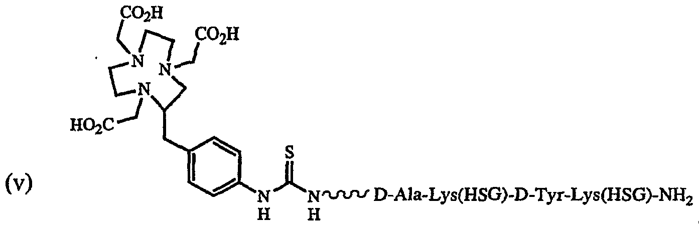


Claims


Priority Applications (11)
| Application Number | Priority Date | Filing Date | Title |
|---|---|---|---|
| CN038192942A CN1675245B (en) | 2002-06-14 | 2003-06-16 | Monoclonal antibody hPAM4 |
| KR1020047020362A KR101143035B1 (en) | 2002-06-14 | 2003-06-16 | Monoclonal antibody hPAM4 |
| ES03740743.4T ES2524767T3 (en) | 2002-06-14 | 2003-06-16 | HPAM4 humanized monoclonal antibody |
| CA2489467A CA2489467C (en) | 2002-06-14 | 2003-06-16 | Humanized monoclonal antibody hpam4 |
| AU2003277087A AU2003277087B2 (en) | 2002-06-14 | 2003-06-16 | Humanized monoclonal antiboby hPAM4 |
| EP03740743.4A EP1519958B1 (en) | 2002-06-14 | 2003-06-16 | Humanized monoclonal antibody hpam4 |
| JP2004513326A JP5139626B2 (en) | 2002-06-14 | 2003-06-16 | Monoclonal antibody hPAM4 |
| MXPA04012656A MXPA04012656A (en) | 2002-06-14 | 2003-06-16 | Humanized monoclonal antiboby hpam4. |
| BR0311799-5A BR0311799A (en) | 2002-06-14 | 2003-06-16 | Hpam4 Monoclonal Antibody |
| IL165753A IL165753A (en) | 2003-06-16 | 2004-12-14 | HUMANIZED MONOCLONAL ANTIBODY hPAM4, A CONJUGATE COMPRISING IT AND AN ANTIBODY COMPRISING THE SAME OR FRAGMENTS THEREOF |
| AU2008212083A AU2008212083B2 (en) | 2002-06-14 | 2008-09-10 | Humanized monoclonal Antibody hPAM4 |
Applications Claiming Priority (2)
| Application Number | Priority Date | Filing Date | Title |
|---|---|---|---|
| US38831402P | 2002-06-14 | 2002-06-14 | |
| US60/388,314 | 2002-06-14 |
Publications (2)
| Publication Number | Publication Date |
|---|---|
| WO2003106495A2 true WO2003106495A2 (en) | 2003-12-24 |
| WO2003106495A3 WO2003106495A3 (en) | 2004-04-01 |
Family
ID=29736456
Family Applications (1)
| Application Number | Title | Priority Date | Filing Date |
|---|---|---|---|
| PCT/GB2003/002593 WO2003106495A2 (en) | 2002-06-14 | 2003-06-16 | MONOCLONAL ANTIBODY hPAM4 |
Country Status (11)
| Country | Link |
|---|---|
| US (2) | US7282567B2 (en) |
| EP (2) | EP1519958B1 (en) |
| JP (2) | JP5139626B2 (en) |
| KR (1) | KR101143035B1 (en) |
| CN (2) | CN1675245B (en) |
| AU (2) | AU2003277087B2 (en) |
| BR (1) | BR0311799A (en) |
| CA (1) | CA2489467C (en) |
| ES (1) | ES2524767T3 (en) |
| MX (1) | MXPA04012656A (en) |
| WO (1) | WO2003106495A2 (en) |
Cited By (31)
| Publication number | Priority date | Publication date | Assignee | Title |
|---|---|---|---|---|
| EP1448780A2 (en) * | 2001-10-15 | 2004-08-25 | Immunomedics, Inc. | Direct targeting binding proteins |
| JP2008528623A (en) * | 2005-01-28 | 2008-07-31 | ラモット アット テル アビブ ユニバーシティ, リミテッド | Α / β antibody of anti-MUC1 |
| WO2011068845A1 (en) * | 2009-12-02 | 2011-06-09 | Immunomedics, Inc. | Combining radioimmunotherapy and antibody-drug conjugates for improved cancer therapy |
| WO2012143496A2 (en) | 2011-04-21 | 2012-10-26 | Bayer Intellectual Property Gmbh | Novel binder-drug conjugates (adcs) and their use |
| WO2012112443A3 (en) * | 2011-02-15 | 2012-11-22 | Immunomedics, Inc. | Anti-mucin antibodies for early detection and treatment of pancreatic cancer |
| US20130039861A1 (en) * | 2005-04-06 | 2013-02-14 | Immunomedics, Inc. | Dye Conjugated Peptides for Fluorescent Imaging |
| US8435529B2 (en) | 2002-06-14 | 2013-05-07 | Immunomedics, Inc. | Combining radioimmunotherapy and antibody-drug conjugates for improved cancer therapy |
| US8859495B2 (en) * | 2005-03-30 | 2014-10-14 | Minerva Biotechnologies Corporation | Methods for stimulating or enhancing proliferation of non-tumorous cells expressing MUC1 receptors |
| US9005613B2 (en) | 2003-06-16 | 2015-04-14 | Immunomedics, Inc. | Anti-mucin antibodies for early detection and treatment of pancreatic cancer |
| US20150218288A1 (en) * | 2005-03-30 | 2015-08-06 | Minerva Biotechnologies Corporation | Proliferation of muc1 expressing cells |
| US9128082B2 (en) | 2009-03-24 | 2015-09-08 | Biocept, Inc. | Devices and methods of cell capture and analysis |
| US9452228B2 (en) | 2013-04-01 | 2016-09-27 | Immunomedics, Inc. | Antibodies reactive with an epitope located in the N-terminal region of MUC5AC comprising cysteine-rich subdomain 2 (Cys2) |
| WO2016207089A1 (en) | 2015-06-22 | 2016-12-29 | Bayer Pharma Aktiengesellschaft | Antibody drug conjugates (adcs) and antibody prodrug conjugates (apdcs) with enzymatically cleavable groups |
| WO2017060322A2 (en) | 2015-10-10 | 2017-04-13 | Bayer Pharma Aktiengesellschaft | Ptefb-inhibitor-adc |
| US9671407B2 (en) | 2009-03-24 | 2017-06-06 | Biocept, Inc. | Devices and methods of cell capture and analysis |
| US9670286B2 (en) | 2012-08-14 | 2017-06-06 | Ibc Pharmaceuticals, Inc. | Disease therapy by inducing immune response to Trop-2 expressing cells |
| WO2017162663A1 (en) | 2016-03-24 | 2017-09-28 | Bayer Pharma Aktiengesellschaft | Prodrugs of cytotoxic active agents having enzymatically cleavable groups |
| US9796788B2 (en) | 2010-02-08 | 2017-10-24 | Regeneron Pharmaceuticals, Inc. | Mice expressing a limited immunoglobulin light chain repertoire |
| US9969814B2 (en) | 2010-02-08 | 2018-05-15 | Regeneron Pharmaceuticals, Inc. | Methods for making fully human bispecific antibodies using a common light chain |
| WO2018114578A1 (en) | 2016-12-21 | 2018-06-28 | Bayer Pharma Aktiengesellschaft | Antibody drug conjugates (adcs) having enzymatically cleavable groups |
| WO2018114798A1 (en) | 2016-12-21 | 2018-06-28 | Bayer Aktiengesellschaft | Prodrugs of cytotoxic active agents having enzymatically cleavable groups |
| US10022453B2 (en) | 2013-12-23 | 2018-07-17 | Bayer Pharma Aktiengesellschaft | Antibody drug conjugates (ADCs) with kinesin spindel protein (KSP) |
| US10130081B2 (en) | 2011-08-05 | 2018-11-20 | Regeneron Pharmaceuticals, Inc. | Humanized universal light chain mice |
| US10143186B2 (en) | 2010-02-08 | 2018-12-04 | Regeneron Pharmaceuticals, Inc. | Common light chain mouse |
| US10881085B2 (en) | 2014-03-21 | 2021-01-05 | Regeneron Pharmaceuticals, Inc. | Non-human animals that make single domain binding proteins |
| EP3643315A4 (en) * | 2017-06-22 | 2021-03-31 | JE&NA Biotech. Co., Ltd. | Composition comprising catalase, preparation method and use thereof and method for killing tumor cells |
| US11001636B2 (en) | 2016-06-15 | 2021-05-11 | Bayer Pharma Aktiengesellschaft | Specific antibody-drug-conjugates (ADCs) with KSP inhibitors and anti-CD123-antibodies |
| US11111314B2 (en) | 2015-03-19 | 2021-09-07 | Regeneron Pharmaceuticals, Inc. | Non-human animals that select for light chain variable regions that bind antigen |
| US11433140B2 (en) | 2016-12-21 | 2022-09-06 | Bayer Pharma Aktiengesellschaft | Specific antibody drug conjugates (ADCs) having KSP inhibitors |
| US11793888B2 (en) | 2015-11-23 | 2023-10-24 | Sangamo Therapeutics, Inc. | Methods and compositions for engineering immunity |
| US11898160B2 (en) | 2008-10-09 | 2024-02-13 | Minerva Biotechnologies Corporation | Method for maintaining pluripotency in cells |
Families Citing this family (82)
| Publication number | Priority date | Publication date | Assignee | Title |
|---|---|---|---|---|
| US7550143B2 (en) | 2005-04-06 | 2009-06-23 | Ibc Pharmaceuticals, Inc. | Methods for generating stably linked complexes composed of homodimers, homotetramers or dimers of dimers and uses |
| US8877901B2 (en) | 2002-12-13 | 2014-11-04 | Immunomedics, Inc. | Camptothecin-binding moiety conjugates |
| US7591994B2 (en) | 2002-12-13 | 2009-09-22 | Immunomedics, Inc. | Camptothecin-binding moiety conjugates |
| US8491896B2 (en) * | 2002-06-14 | 2013-07-23 | Immunomedics, Inc. | Anti-pancreatic cancer antibodies |
| US20160279239A1 (en) | 2011-05-02 | 2016-09-29 | Immunomedics, Inc. | Subcutaneous administration of anti-cd74 antibody for systemic lupus erythematosus and autoimmune disease |
| US8821868B2 (en) | 2002-06-14 | 2014-09-02 | Immunomedics, Inc. | Anti-pancreatic cancer antibodies |
| US9599619B2 (en) | 2002-06-14 | 2017-03-21 | Immunomedics, Inc. | Anti-pancreatic cancer antibodies |
| US7902338B2 (en) | 2003-07-31 | 2011-03-08 | Immunomedics, Inc. | Anti-CD19 antibodies |
| AU2006218454B2 (en) | 2005-03-03 | 2011-11-17 | Immunomedics, Inc. | Humanized L243 antibodies |
| US20160355591A1 (en) | 2011-05-02 | 2016-12-08 | Immunomedics, Inc. | Subcutaneous anti-hla-dr monoclonal antibody for treatment of hematologic malignancies |
| JP2006248978A (en) * | 2005-03-10 | 2006-09-21 | Mebiopharm Co Ltd | New liposome preparation |
| EP2674440B1 (en) | 2005-12-16 | 2019-07-03 | IBC Pharmaceuticals, Inc. | Multivalent immunoglobulin-based bioactive assemblies |
| DK1991273T4 (en) * | 2006-02-10 | 2022-02-07 | Life Technologies Corp | LABELING AND DETECTION OF POST-TRANSLATIONALLY MODIFIED PROTEINS |
| SI2423230T1 (en) * | 2006-03-27 | 2013-09-30 | Medimmune Limited | Binding member for GM-CSF receptor |
| JP2010505775A (en) | 2006-10-04 | 2010-02-25 | クーベンハヴンス・ユニヴェルシテット | Production of cancer-specific immune response against MUC1 and cancer-specific MUC1 antibody |
| US7777877B2 (en) * | 2006-10-20 | 2010-08-17 | California Institute Of Technology | High efficiency coupling optics for pumping and detection of fluorescence |
| TWI501976B (en) * | 2007-05-21 | 2015-10-01 | Alder Biopharmaceuticals Inc | Antibodies to tnf alpha and use thereof |
| US8252286B2 (en) | 2007-05-21 | 2012-08-28 | Alderbio Holdings Llc | Antagonists of IL-6 to prevent or treat thrombosis |
| US8062864B2 (en) | 2007-05-21 | 2011-11-22 | Alderbio Holdings Llc | Nucleic acids encoding antibodies to IL-6, and recombinant production of anti-IL-6 antibodies |
| US8404235B2 (en) * | 2007-05-21 | 2013-03-26 | Alderbio Holdings Llc | Antagonists of IL-6 to raise albumin and/or lower CRP |
| US8178101B2 (en) | 2007-05-21 | 2012-05-15 | Alderbio Holdings Inc. | Use of anti-IL-6 antibodies having specific binding properties to treat cachexia |
| TWI561530B (en) | 2007-05-21 | 2016-12-11 | Alderbio Holdings Llc | Antibodies to il-6 and use thereof |
| US9701747B2 (en) | 2007-05-21 | 2017-07-11 | Alderbio Holdings Llc | Method of improving patient survivability and quality of life by anti-IL-6 antibody administration |
| US20090238825A1 (en) * | 2007-05-21 | 2009-09-24 | Kovacevich Brian R | Novel rabbit antibody humanization methods and humanized rabbit antibodies |
| US7906117B2 (en) | 2007-05-21 | 2011-03-15 | Alderbio Holdings Llc | Antagonists of IL-6 to prevent or treat cachexia, weakness, fatigue, and/or fever |
| WO2009142460A2 (en) * | 2008-05-23 | 2009-11-26 | Samsung Electronics Co., Ltd. | Antibody-peptide fused synergibody |
| US8489177B2 (en) * | 2008-07-16 | 2013-07-16 | Dilon Technologies, Inc. | Fiducial marker and method for gamma guided stereotactic localization |
| JP5919604B2 (en) * | 2008-08-08 | 2016-05-18 | イミューノメディクス、インコーポレイテッドImmunomedics, Inc. | Anti-pancreatic cancer antibody |
| EP4083072A1 (en) * | 2008-10-06 | 2022-11-02 | Minerva Biotechnologies Corporation | Muc1* antibodies |
| US9452227B2 (en) | 2008-11-25 | 2016-09-27 | Alderbio Holdings Llc | Methods of treating or diagnosing conditions associated with elevated IL-6 using anti-IL-6 antibodies or fragments |
| US8337847B2 (en) | 2008-11-25 | 2012-12-25 | Alderbio Holdings Llc | Methods of treating anemia using anti-IL-6 antibodies |
| US8992920B2 (en) | 2008-11-25 | 2015-03-31 | Alderbio Holdings Llc | Anti-IL-6 antibodies for the treatment of arthritis |
| US8420089B2 (en) | 2008-11-25 | 2013-04-16 | Alderbio Holdings Llc | Antagonists of IL-6 to raise albumin and/or lower CRP |
| US8323649B2 (en) | 2008-11-25 | 2012-12-04 | Alderbio Holdings Llc | Antibodies to IL-6 and use thereof |
| US9212223B2 (en) | 2008-11-25 | 2015-12-15 | Alderbio Holdings Llc | Antagonists of IL-6 to prevent or treat thrombosis |
| DK3903829T3 (en) | 2009-02-13 | 2023-06-26 | Immunomedics Inc | IMMUNE CONJUGATES WITH AN INTRACELLULAR CLEAVABLE BOND |
| IN2012DN01663A (en) | 2009-09-16 | 2015-06-05 | Immunomedics Inc | |
| US8518405B2 (en) * | 2009-10-08 | 2013-08-27 | The University Of North Carolina At Charlotte | Tumor specific antibodies and uses therefor |
| EP2504031A4 (en) | 2009-11-24 | 2013-06-26 | Alderbio Holdings Llc | Antibodies to il-6 and use thereof |
| US9775921B2 (en) | 2009-11-24 | 2017-10-03 | Alderbio Holdings Llc | Subcutaneously administrable composition containing anti-IL-6 antibody |
| CA2787753A1 (en) * | 2010-01-22 | 2011-07-28 | Immunomedics, Inc. | Detection of early-stage pancreatic adenocarcinoma |
| US20120021409A1 (en) * | 2010-02-08 | 2012-01-26 | Regeneron Pharmaceuticals, Inc. | Common Light Chain Mouse |
| US9845362B2 (en) | 2010-10-08 | 2017-12-19 | The University Of North Carolina At Charlotte | Compositions comprising chimeric antigen receptors, T cells comprising the same, and methods of using the same |
| CA2818813C (en) | 2010-11-23 | 2020-10-06 | Alder Biopharmaceuticals, Inc. | Anti-il-6 antibodies for the treatment of oral mucositis |
| CA2831572C (en) | 2011-05-02 | 2019-11-26 | Immunomedics, Inc. | Ultrafiltration concentration of allotype selected antibodies for small-volume administration |
| KR20150006000A (en) | 2012-05-01 | 2015-01-15 | 제넨테크, 인크. | Anti-pmel17 antibodies and immunoconjugates |
| EP2854523A1 (en) * | 2012-06-05 | 2015-04-08 | Regeneron Pharmaceuticals, Inc. | Methods for making fully human bispecific antibodies using a common light chain |
| CA2874864C (en) | 2012-08-14 | 2023-02-21 | Ibc Pharmaceuticals, Inc. | T-cell redirecting bispecific antibodies for treatment of disease |
| WO2017004144A1 (en) | 2015-07-01 | 2017-01-05 | Immunomedics, Inc. | Antibody-sn-38 immunoconjugates with a cl2a linker |
| HUE057977T2 (en) | 2012-12-13 | 2022-06-28 | Immunomedics Inc | Dosages of immunoconjugates of antibodies and sn-38 for improved efficacy and decreased toxicity |
| US9989524B2 (en) | 2013-02-05 | 2018-06-05 | Sanofi | Immuno imaging agent for use with antibody-drug conjugate therapy |
| JP2016515093A (en) * | 2013-02-05 | 2016-05-26 | サノフイ | Immunoimaging agents for use with antibody-drug conjugate therapy |
| EP2774930A1 (en) | 2013-03-07 | 2014-09-10 | Aptenia S.R.L. | Metallocene compounds and labeled molecules comprising the same for in vivo imaging. |
| CA2899811A1 (en) * | 2013-04-01 | 2014-10-09 | Immunomedics, Inc. | Anti-mucin antibodies for early detection and treament of pancreatic cancer |
| WO2014198995A1 (en) * | 2013-06-14 | 2014-12-18 | Universidad De Granada | Biomarkers for the diagnosis and the response to treatment of pancreatic cancer |
| TN2016000137A1 (en) | 2013-10-18 | 2017-10-06 | Deutsches Krebsforsch | Labeled inhibitors of prostate specific membrane antigen (psma), their use as imaging agents and pharmaceutical agents for the treatment of prostate cancer. |
| MX2016010683A (en) | 2014-02-21 | 2017-05-11 | Ibc Pharmaceuticals Inc | Disease therapy by inducing immune response to trop-2 expressing cells. |
| US9139649B2 (en) | 2014-02-25 | 2015-09-22 | Immunomedics, Inc. | Humanized anti-CD22 antibody |
| EP3126383B8 (en) | 2014-04-03 | 2019-02-27 | IGM Biosciences, Inc. | Modified j-chain |
| GB201406767D0 (en) | 2014-04-15 | 2014-05-28 | Cancer Rec Tech Ltd | Humanized anti-Tn-MUC1 antibodies anf their conjugates |
| WO2015200260A1 (en) | 2014-06-24 | 2015-12-30 | Immunomedics, Inc. | Anti-histone therapy for vascular necrosis in severe glomerulonephritis |
| CN105334207A (en) * | 2014-08-15 | 2016-02-17 | 上海中信国健药业股份有限公司 | Biological activity detection method by aiming at EGFR targeted therapeutic medicine |
| US11554181B2 (en) | 2014-09-05 | 2023-01-17 | The University Of North Carolina At Charlotte | Tumor specific antibody conjugates and uses therefor |
| JP7080055B2 (en) | 2014-09-08 | 2022-06-03 | ルタナ ゲーエムベーハー | Constructs for delivery of molecules to the cytoplasm of cells |
| PL3204018T3 (en) | 2014-10-07 | 2022-01-03 | Immunomedics, Inc. | Neoadjuvant use of antibody-drug conjugates |
| IL297997A (en) | 2015-03-04 | 2023-01-01 | Igm Biosciences Inc | Cd20 binding molecules and uses thereof |
| CA2981543A1 (en) | 2015-04-22 | 2016-10-27 | Immunomedics, Inc. | Isolation, detection, diagnosis and/or characterization of circulating trop-2-positive cancer cells |
| WO2016176332A1 (en) * | 2015-04-27 | 2016-11-03 | The Regents Of The University Of Colorado | Anthracycline prodrugs and methods of making and using the same |
| ES2953441T3 (en) | 2015-06-25 | 2023-11-13 | Immunomedics Inc | Combination of anti-hla-dr or anti-Trop-2 antibodies with microtubule inhibitors, parp inhibitors, bruton kinase inhibitors or phosphoinositide 3-kinase inhibitors significantly improves therapeutic outcome in cancer |
| CA2999284C (en) | 2015-09-30 | 2023-06-13 | Igm Biosciences A/S | Binding molecules with modified j-chain |
| DK3356401T3 (en) | 2015-09-30 | 2020-09-07 | Igm Biosciences Inc | BINDING MOLECULES WITH MODIFIED J-CHAIN |
| EP3606964A4 (en) | 2017-04-03 | 2020-12-09 | Immunomedics, Inc. | Subcutaneous administration of antibody-drug conjugates for cancer therapy |
| CN111542324B (en) * | 2018-02-11 | 2023-09-12 | 四川科伦博泰生物医药股份有限公司 | Cytotoxic agent and conjugate thereof, preparation method and application thereof |
| AU2019348440A1 (en) | 2018-09-26 | 2021-03-25 | Ascendis Pharma A/S | Degradable hyaluronic acid hydrogels |
| CN111362926B (en) * | 2018-12-26 | 2021-04-30 | 联宁(苏州)生物制药有限公司 | Synthetic method of intermediate CLA-SN38 for antibody coupled drug and intermediate thereof |
| WO2022010797A2 (en) | 2020-07-07 | 2022-01-13 | Bionecure Therapeutics, Inc. | Novel maytansinoids as adc payloads and their use for the treatment of cancer |
| CN115803010A (en) | 2020-07-09 | 2023-03-14 | 豪夫迈·罗氏有限公司 | Concentrated protein compositions, their preparation and use |
| CN115212308B (en) * | 2021-04-15 | 2023-10-20 | 中国医学科学院基础医学研究所 | Application of GASDERMIN E pathway targeting agent in treating pancreatic cancer |
| TW202308706A (en) * | 2021-04-21 | 2023-03-01 | 日商日本醫事物理股份有限公司 | Radioactive antitumor agent |
| JPWO2022239720A1 (en) | 2021-05-10 | 2022-11-17 | ||
| WO2023016488A1 (en) | 2021-08-13 | 2023-02-16 | 昆山新蕴达生物科技有限公司 | Microtubule inhibitor-based antibody-drug conjugate |
| WO2023024949A1 (en) | 2021-08-24 | 2023-03-02 | 昆山新蕴达生物科技有限公司 | Antibody-drug conjugate conjugated via breakable linker |
Family Cites Families (45)
| Publication number | Priority date | Publication date | Assignee | Title |
|---|---|---|---|---|
| US4036945A (en) | 1976-05-03 | 1977-07-19 | The Massachusetts General Hospital | Composition and method for determining the size and location of myocardial infarcts |
| US4331647A (en) | 1980-03-03 | 1982-05-25 | Goldenberg Milton David | Tumor localization and therapy with labeled antibody fragments specific to tumor-associated markers |
| US4671958A (en) | 1982-03-09 | 1987-06-09 | Cytogen Corporation | Antibody conjugates for the delivery of compounds to target sites |
| US4624846A (en) | 1983-07-29 | 1986-11-25 | Immunomedics, Inc. | Method for enhancing target specificity of antibody localization and clearance of non-target diagnostic and therapeutic principles |
| US4824659A (en) | 1985-06-07 | 1989-04-25 | Immunomedics, Inc. | Antibody conjugates |
| US5776093A (en) | 1985-07-05 | 1998-07-07 | Immunomedics, Inc. | Method for imaging and treating organs and tissues |
| US5057313A (en) | 1986-02-25 | 1991-10-15 | The Center For Molecular Medicine And Immunology | Diagnostic and therapeutic antibody conjugates |
| US4699784A (en) | 1986-02-25 | 1987-10-13 | Center For Molecular Medicine & Immunology | Tumoricidal methotrexate-antibody conjugate |
| US4704692A (en) | 1986-09-02 | 1987-11-03 | Ladner Robert C | Computer based system and method for determining and displaying possible chemical structures for converting double- or multiple-chain polypeptides to single-chain polypeptides |
| US4946778A (en) | 1987-09-21 | 1990-08-07 | Genex Corporation | Single polypeptide chain binding molecules |
| US5567610A (en) | 1986-09-04 | 1996-10-22 | Bioinvent International Ab | Method of producing human monoclonal antibodies and kit therefor |
| FR2604092B1 (en) | 1986-09-19 | 1990-04-13 | Immunotech Sa | IMMUNOREACTIVES FOR TARGETING ANIMAL CELLS FOR VISUALIZATION OR DESTRUCTION IN VIVO |
| US5019368A (en) | 1989-02-23 | 1991-05-28 | Cancer Biologics, Inc. | Detection of necrotic malignant tissue and associated therapy |
| US5132405A (en) | 1987-05-21 | 1992-07-21 | Creative Biomolecules, Inc. | Biosynthetic antibody binding sites |
| US4925648A (en) | 1988-07-29 | 1990-05-15 | Immunomedics, Inc. | Detection and treatment of infectious and inflammatory lesions |
| US5734033A (en) | 1988-12-22 | 1998-03-31 | The Trustees Of The University Of Pennsylvania | Antisense oligonucleotides inhibiting human bcl-2 gene expression |
| US5530101A (en) * | 1988-12-28 | 1996-06-25 | Protein Design Labs, Inc. | Humanized immunoglobulins |
| US4935498A (en) * | 1989-03-06 | 1990-06-19 | Board Of Regents, The University Of Texas System | Expanded porphyrins: large porphyrin-like tripyrroledimethine-derived macrocycles |
| US5859205A (en) * | 1989-12-21 | 1999-01-12 | Celltech Limited | Humanised antibodies |
| US5229275A (en) | 1990-04-26 | 1993-07-20 | Akzo N.V. | In-vitro method for producing antigen-specific human monoclonal antibodies |
| US5633425A (en) | 1990-08-29 | 1997-05-27 | Genpharm International, Inc. | Transgenic non-human animals capable of producing heterologous antibodies |
| US5660827A (en) * | 1992-03-05 | 1997-08-26 | Board Of Regents, The University Of Texas System | Antibodies that bind to endoglin |
| ATE239506T1 (en) | 1992-03-05 | 2003-05-15 | Univ Texas | USE OF IMMUNOCONJUGATES FOR THE DIAGNOSIS AND/OR THERAPY OF VASCULARIZED TUMORS |
| US5965132A (en) | 1992-03-05 | 1999-10-12 | Board Of Regents, The University Of Texas System | Methods and compositions for targeting the vasculature of solid tumors |
| US6096289A (en) | 1992-05-06 | 2000-08-01 | Immunomedics, Inc. | Intraoperative, intravascular, and endoscopic tumor and lesion detection, biopsy and therapy |
| ATE194916T1 (en) | 1992-05-06 | 2000-08-15 | Immunomedics Inc | INTRAOPERATIVE, INTRAVASCULAR AND ENDOSCOPIC DETERMINATION AND TREATMENT OF INJURIES AND TUMORS |
| EP1550729B1 (en) | 1992-09-25 | 2009-05-27 | Avipep Pty Limited | Target binding polypeptide comprising an IG-like VL domain linked to an IG-like VH domain |
| US5837242A (en) | 1992-12-04 | 1998-11-17 | Medical Research Council | Multivalent and multispecific binding proteins, their manufacture and use |
| US5443953A (en) | 1993-12-08 | 1995-08-22 | Immunomedics, Inc. | Preparation and use of immunoconjugates |
| EP0771208B1 (en) * | 1994-08-12 | 2005-10-19 | Immunomedics, Inc. | Immunoconjugates and humanized antibodies specific for b-cell lymphoma and leukemia cells |
| US6176842B1 (en) * | 1995-03-08 | 2001-01-23 | Ekos Corporation | Ultrasound assembly for use with light activated drugs |
| AUPO591797A0 (en) | 1997-03-27 | 1997-04-24 | Commonwealth Scientific And Industrial Research Organisation | High avidity polyvalent and polyspecific reagents |
| CA2222140C (en) | 1995-06-15 | 2010-11-23 | Introgene B.V. | Packaging systems for human recombinant adenovirus to be used in gene therapy |
| WO1997034632A1 (en) | 1996-03-20 | 1997-09-25 | Immunomedics, Inc. | Glycosylated humanized b-cell specific antibodies |
| EP0954340B1 (en) | 1996-05-03 | 2007-06-27 | Immunomedics, Inc. | Targeted combination immunotherapy of cancer |
| US6261537B1 (en) * | 1996-10-28 | 2001-07-17 | Nycomed Imaging As | Diagnostic/therapeutic agents having microbubbles coupled to one or more vectors |
| US6306393B1 (en) * | 1997-03-24 | 2001-10-23 | Immunomedics, Inc. | Immunotherapy of B-cell malignancies using anti-CD22 antibodies |
| US6120995A (en) * | 1997-08-07 | 2000-09-19 | Thomas Jefferson University | Compositions that specifically bind to colorectal cancer cells and methods of using the same |
| US7138103B2 (en) * | 1998-06-22 | 2006-11-21 | Immunomedics, Inc. | Use of bi-specific antibodies for pre-targeting diagnosis and therapy |
| US7405320B2 (en) * | 1998-06-22 | 2008-07-29 | Immunomedics, Inc. | Therapeutic and diagnostic conjugates for use with multispecific antibodies |
| EP1135498B1 (en) * | 1998-11-18 | 2008-01-23 | Genentech, Inc. | Antibody variants with higher binding affinity compared to parent antibodies |
| WO2000063403A2 (en) | 1999-04-15 | 2000-10-26 | Crucell Holland B.V. | Recombinant protein production in a human cell using sequences encoding adenovirus e1 protein |
| ES2223705T3 (en) | 1999-04-28 | 2005-03-01 | Board Of Regents, The University Of Texas System | COMPOSITIONS AND METHODS FOR THE TREATMENT OF CANCER THROUGH INHIBITION SELECTIVE OF VEGF. |
| AU2001297625A1 (en) * | 2000-12-21 | 2002-09-12 | Bristol-Myers Squibb Company | Nucleic acid molecules and polypeptides for a human cation channel polypeptide |
| AU2002360766A1 (en) * | 2001-12-21 | 2003-07-30 | Threshold Pharmaceuticals, Inc. | Methods for cancer imaging |
-
2003
- 2003-06-16 CN CN038192942A patent/CN1675245B/en not_active Expired - Fee Related
- 2003-06-16 US US10/461,885 patent/US7282567B2/en not_active Expired - Fee Related
- 2003-06-16 EP EP03740743.4A patent/EP1519958B1/en not_active Expired - Lifetime
- 2003-06-16 KR KR1020047020362A patent/KR101143035B1/en not_active IP Right Cessation
- 2003-06-16 ES ES03740743.4T patent/ES2524767T3/en not_active Expired - Lifetime
- 2003-06-16 AU AU2003277087A patent/AU2003277087B2/en not_active Ceased
- 2003-06-16 CA CA2489467A patent/CA2489467C/en not_active Expired - Fee Related
- 2003-06-16 CN CN2010101460004A patent/CN101824090B/en not_active Expired - Fee Related
- 2003-06-16 WO PCT/GB2003/002593 patent/WO2003106495A2/en active Application Filing
- 2003-06-16 JP JP2004513326A patent/JP5139626B2/en not_active Expired - Fee Related
- 2003-06-16 BR BR0311799-5A patent/BR0311799A/en not_active Application Discontinuation
- 2003-06-16 MX MXPA04012656A patent/MXPA04012656A/en active IP Right Grant
- 2003-06-16 EP EP10012295A patent/EP2301968A3/en not_active Ceased
-
2007
- 2007-09-04 US US11/849,791 patent/US20080050311A1/en not_active Abandoned
-
2008
- 2008-09-10 AU AU2008212083A patent/AU2008212083B2/en not_active Ceased
-
2010
- 2010-04-13 JP JP2010092200A patent/JP5155356B2/en not_active Expired - Fee Related
Non-Patent Citations (5)
| Title |
|---|
| CARDILLO T M ET AL., CLINICAL CANCER RESEARCH, vol. 7, October 2001 (2001-10-01), pages 3186 - 3192 |
| CARDILLO T M ET AL., INTERNATIONAL JOURNAL OF CANCER, vol. 97, March 2002 (2002-03-01), pages 386 - 392 |
| GOLD D V ET AL., PROCEEDINGS OF THE AMERICAN ASSOCIATION FOR CANCER RESEARCH ANNUAL MEETING, vol. 34, 19 May 1993 (1993-05-19), pages 480 |
| GOLD D V ET AL., THE INTERNATIONAL JOURNAL OF CANCER, vol. 71, 1997, pages 660 - 667 |
| MARIANI G ET AL., CANCER RESEARCH (SUPPL, vol. 55, December 1995 (1995-12-01), pages 5911S - 5915S |
Cited By (57)
| Publication number | Priority date | Publication date | Assignee | Title |
|---|---|---|---|---|
| EP1448780A4 (en) * | 2001-10-15 | 2005-08-31 | Immunomedics Inc | Direct targeting binding proteins |
| EP1448780A2 (en) * | 2001-10-15 | 2004-08-25 | Immunomedics, Inc. | Direct targeting binding proteins |
| US9272057B2 (en) | 2002-02-26 | 2016-03-01 | Immunomedics, Inc. | Combining radioimmunotherapy and antibody-drug conjugates for improved cancer therapy |
| US8992931B2 (en) | 2002-03-01 | 2015-03-31 | Immunomedics, Inc. | Combining radioimmunotherapy and antibody-drug conjugates for improved cancer therapy |
| US8574854B2 (en) | 2002-06-14 | 2013-11-05 | Immunomedics, Inc. | Anti-mucin antibodies for early detection and treatment of pancreatic cancer |
| US9089618B2 (en) | 2002-06-14 | 2015-07-28 | Immunomedics, Inc. | Anti-mucin antibodies for early detection and treatment of pancreatic cancer |
| US8795662B2 (en) | 2002-06-14 | 2014-08-05 | Immunomedics, Inc. | Anti-mucin antibodies for early detection and treatment of pancreatic cancer |
| US8586050B2 (en) | 2002-06-14 | 2013-11-19 | Immunomedics, Inc. | Combining radioimmunotherapy and antibody-drug conjugates for improved cancer therapy |
| US8435529B2 (en) | 2002-06-14 | 2013-05-07 | Immunomedics, Inc. | Combining radioimmunotherapy and antibody-drug conjugates for improved cancer therapy |
| US9005613B2 (en) | 2003-06-16 | 2015-04-14 | Immunomedics, Inc. | Anti-mucin antibodies for early detection and treatment of pancreatic cancer |
| US9238084B2 (en) | 2003-06-16 | 2016-01-19 | Immunomedics, Inc. | Anti-mucin antibodies for early detection and treatment of pancreatic cancer |
| JP2008528623A (en) * | 2005-01-28 | 2008-07-31 | ラモット アット テル アビブ ユニバーシティ, リミテッド | Α / β antibody of anti-MUC1 |
| US10703821B2 (en) | 2005-03-30 | 2020-07-07 | Minerva Biotechnologies Corporation | Method for stimulating or enhancing proliferation of cells by activating the mucin 1 (MUC1) receptor |
| US8859495B2 (en) * | 2005-03-30 | 2014-10-14 | Minerva Biotechnologies Corporation | Methods for stimulating or enhancing proliferation of non-tumorous cells expressing MUC1 receptors |
| US20150218288A1 (en) * | 2005-03-30 | 2015-08-06 | Minerva Biotechnologies Corporation | Proliferation of muc1 expressing cells |
| US20130039861A1 (en) * | 2005-04-06 | 2013-02-14 | Immunomedics, Inc. | Dye Conjugated Peptides for Fluorescent Imaging |
| US11898160B2 (en) | 2008-10-09 | 2024-02-13 | Minerva Biotechnologies Corporation | Method for maintaining pluripotency in cells |
| US10527611B2 (en) | 2009-03-24 | 2020-01-07 | Biocept, Inc. | Devices and methods of cell capture and analysis |
| US11719692B2 (en) | 2009-03-24 | 2023-08-08 | Biocept, Inc. | Devices and methods of cell capture and analysis |
| US9671407B2 (en) | 2009-03-24 | 2017-06-06 | Biocept, Inc. | Devices and methods of cell capture and analysis |
| US9128082B2 (en) | 2009-03-24 | 2015-09-08 | Biocept, Inc. | Devices and methods of cell capture and analysis |
| WO2011068845A1 (en) * | 2009-12-02 | 2011-06-09 | Immunomedics, Inc. | Combining radioimmunotherapy and antibody-drug conjugates for improved cancer therapy |
| US9969814B2 (en) | 2010-02-08 | 2018-05-15 | Regeneron Pharmaceuticals, Inc. | Methods for making fully human bispecific antibodies using a common light chain |
| US11026407B2 (en) | 2010-02-08 | 2021-06-08 | Regeneran Pharmaceuticals, Inc. | Mice expressing a limited immunoglobulin light chain repertoire |
| US10412940B2 (en) | 2010-02-08 | 2019-09-17 | Regeneron Pharmaceuticals, Inc. | Mice expressing a limited immunoglobulin light chain repertoire |
| US10167344B2 (en) | 2010-02-08 | 2019-01-01 | Regeneron Pharmaceuticals, Inc. | Mice expressing a limited immunoglobulin light chain repertoire |
| US10143186B2 (en) | 2010-02-08 | 2018-12-04 | Regeneron Pharmaceuticals, Inc. | Common light chain mouse |
| US9796788B2 (en) | 2010-02-08 | 2017-10-24 | Regeneron Pharmaceuticals, Inc. | Mice expressing a limited immunoglobulin light chain repertoire |
| US10986820B2 (en) | 2010-02-08 | 2021-04-27 | Regeneron Pharmaceuticals, Inc. | Common light chain mouse |
| WO2012112443A3 (en) * | 2011-02-15 | 2012-11-22 | Immunomedics, Inc. | Anti-mucin antibodies for early detection and treatment of pancreatic cancer |
| CN103429263A (en) * | 2011-02-15 | 2013-12-04 | 免疫医疗公司 | Anti-mucin antibodies for early detection and treatment of pancreatic cancer |
| WO2012143496A2 (en) | 2011-04-21 | 2012-10-26 | Bayer Intellectual Property Gmbh | Novel binder-drug conjugates (adcs) and their use |
| WO2012143499A2 (en) | 2011-04-21 | 2012-10-26 | Bayer Intellectual Property Gmbh | Novel binder-drug conjugates (adcs) and their use |
| US10130081B2 (en) | 2011-08-05 | 2018-11-20 | Regeneron Pharmaceuticals, Inc. | Humanized universal light chain mice |
| US11357217B2 (en) | 2011-08-05 | 2022-06-14 | Regeneron Pharmaceuticals, Inc. | Humanized universal light chain mice |
| US9879088B2 (en) | 2012-08-14 | 2018-01-30 | Ibc Pharmaceuticals, Inc. | Disease therapy by inducing immune response to Trop-2 expressing cells |
| US10662252B2 (en) | 2012-08-14 | 2020-05-26 | Ibc Pharmaceuticals, Inc. | Disease therapy by inducing immune response to Trop-2 expressing cells |
| US9670286B2 (en) | 2012-08-14 | 2017-06-06 | Ibc Pharmaceuticals, Inc. | Disease therapy by inducing immune response to Trop-2 expressing cells |
| US9452228B2 (en) | 2013-04-01 | 2016-09-27 | Immunomedics, Inc. | Antibodies reactive with an epitope located in the N-terminal region of MUC5AC comprising cysteine-rich subdomain 2 (Cys2) |
| US10022453B2 (en) | 2013-12-23 | 2018-07-17 | Bayer Pharma Aktiengesellschaft | Antibody drug conjugates (ADCs) with kinesin spindel protein (KSP) |
| US10881085B2 (en) | 2014-03-21 | 2021-01-05 | Regeneron Pharmaceuticals, Inc. | Non-human animals that make single domain binding proteins |
| US11111314B2 (en) | 2015-03-19 | 2021-09-07 | Regeneron Pharmaceuticals, Inc. | Non-human animals that select for light chain variable regions that bind antigen |
| US11123439B2 (en) | 2015-06-22 | 2021-09-21 | Bayer Pharma Aktiengesellschaft | Antibody drug conjugates (ADCS) and antibody prodrug conjugates (APDCS) with enzymatically cleavable groups |
| WO2016207089A1 (en) | 2015-06-22 | 2016-12-29 | Bayer Pharma Aktiengesellschaft | Antibody drug conjugates (adcs) and antibody prodrug conjugates (apdcs) with enzymatically cleavable groups |
| WO2017060322A2 (en) | 2015-10-10 | 2017-04-13 | Bayer Pharma Aktiengesellschaft | Ptefb-inhibitor-adc |
| US11793888B2 (en) | 2015-11-23 | 2023-10-24 | Sangamo Therapeutics, Inc. | Methods and compositions for engineering immunity |
| WO2017162663A1 (en) | 2016-03-24 | 2017-09-28 | Bayer Pharma Aktiengesellschaft | Prodrugs of cytotoxic active agents having enzymatically cleavable groups |
| US11685714B2 (en) | 2016-03-24 | 2023-06-27 | Bayer Pharma Aktiengesellschaft | Prodrugs of cytotoxic active agents having enzymatically cleavable groups |
| US11001636B2 (en) | 2016-06-15 | 2021-05-11 | Bayer Pharma Aktiengesellschaft | Specific antibody-drug-conjugates (ADCs) with KSP inhibitors and anti-CD123-antibodies |
| US11643469B2 (en) | 2016-06-15 | 2023-05-09 | Bayer Pharma Aktiengesellschaft | Specific antibody-drug-conjugates (ADCs) with KSP inhibitors and anti-CD123-antibodies |
| WO2018114798A1 (en) | 2016-12-21 | 2018-06-28 | Bayer Aktiengesellschaft | Prodrugs of cytotoxic active agents having enzymatically cleavable groups |
| US11478554B2 (en) | 2016-12-21 | 2022-10-25 | Bayer Pharma Aktiengesellschaft | Antibody drug conjugates (ADCS) having enzymatically cleavable groups |
| US11660351B2 (en) | 2016-12-21 | 2023-05-30 | Bayer Aktiengesellschaft | Antibody drug conjugates (ADCs) having enzymatically cleavable groups |
| US11433140B2 (en) | 2016-12-21 | 2022-09-06 | Bayer Pharma Aktiengesellschaft | Specific antibody drug conjugates (ADCs) having KSP inhibitors |
| WO2018114578A1 (en) | 2016-12-21 | 2018-06-28 | Bayer Pharma Aktiengesellschaft | Antibody drug conjugates (adcs) having enzymatically cleavable groups |
| US11173220B2 (en) | 2017-06-22 | 2021-11-16 | Je & Na Biotech. Co., Ltd. | Composition comprising catalase, preparation method and use thereof and method for killing tumor cells |
| EP3643315A4 (en) * | 2017-06-22 | 2021-03-31 | JE&NA Biotech. Co., Ltd. | Composition comprising catalase, preparation method and use thereof and method for killing tumor cells |
Also Published As
| Publication number | Publication date |
|---|---|
| JP2010227099A (en) | 2010-10-14 |
| AU2008212083B2 (en) | 2011-06-09 |
| KR101143035B1 (en) | 2012-05-08 |
| JP5139626B2 (en) | 2013-02-06 |
| AU2008212083A1 (en) | 2008-10-02 |
| EP2301968A2 (en) | 2011-03-30 |
| AU2003277087A1 (en) | 2003-12-31 |
| CA2489467C (en) | 2015-02-24 |
| EP1519958B1 (en) | 2014-10-15 |
| CA2489467A1 (en) | 2003-12-24 |
| MXPA04012656A (en) | 2005-08-15 |
| BR0311799A (en) | 2005-05-10 |
| US20050014207A1 (en) | 2005-01-20 |
| CN1675245B (en) | 2011-01-12 |
| WO2003106495A3 (en) | 2004-04-01 |
| US7282567B2 (en) | 2007-10-16 |
| CN1675245A (en) | 2005-09-28 |
| KR20050010054A (en) | 2005-01-26 |
| JP5155356B2 (en) | 2013-03-06 |
| CN101824090B (en) | 2013-01-02 |
| AU2003277087B2 (en) | 2008-07-31 |
| EP1519958A2 (en) | 2005-04-06 |
| US20080050311A1 (en) | 2008-02-28 |
| CN101824090A (en) | 2010-09-08 |
| ES2524767T3 (en) | 2014-12-12 |
| JP2006513695A (en) | 2006-04-27 |
| EP2301968A3 (en) | 2011-06-29 |
Similar Documents
| Publication | Publication Date | Title |
|---|---|---|
| EP1519958B1 (en) | Humanized monoclonal antibody hpam4 | |
| US7238786B2 (en) | Monoclonal antibody cPAM4 | |
| US8017736B2 (en) | Alpha-fetoprotein Immu31 antibodies and fusion proteins and methods of use thereof | |
| US20090185974A1 (en) | Chimeric, Human and Humanized Anti-Granulocyte Antibodies and Methods of Use | |
| IL165752A (en) | Chimeric monoclonal antibody pam4, a conjugate comprising it and an antibody comprising the same or fragments thereof |
Legal Events
| Date | Code | Title | Description |
|---|---|---|---|
| AK | Designated states |
Kind code of ref document: A2 Designated state(s): AE AG AL AM AT AU AZ BA BB BG BR BY BZ CA CH CN CO CR CU CZ DE DK DM DZ EC EE ES FI GB GD GE GH GM HR HU ID IL IN IS JP KE KG KP KR KZ LC LK LR LS LT LU LV MA MD MG MK MN MW MX MZ NI NO NZ OM PG PH PL PT RO RU SC SD SE SG SK SL TJ TM TN TR TT TZ UA UG US UZ VC VN YU ZA ZM ZW |
|
| AL | Designated countries for regional patents |
Kind code of ref document: A2 Designated state(s): GH GM KE LS MW MZ SD SL SZ TZ UG ZM ZW AM AZ BY KG KZ MD RU TJ TM AT BE BG CH CY CZ DE DK EE ES FI FR GB GR HU IE IT LU MC NL PT RO SE SI SK TR BF BJ CF CG CI CM GA GN GQ GW ML MR NE SN TD TG |
|
| DFPE | Request for preliminary examination filed prior to expiration of 19th month from priority date (pct application filed before 20040101) | ||
| 121 | Ep: the epo has been informed by wipo that ep was designated in this application | ||
| WWE | Wipo information: entry into national phase |
Ref document number: PA/a/2004/012656 Country of ref document: MX Ref document number: 2489467 Country of ref document: CA Ref document number: 2004513326 Country of ref document: JP Ref document number: 1020047020362 Country of ref document: KR |
|
| WWE | Wipo information: entry into national phase |
Ref document number: 2003740743 Country of ref document: EP |
|
| WWP | Wipo information: published in national office |
Ref document number: 1020047020362 Country of ref document: KR |
|
| WWE | Wipo information: entry into national phase |
Ref document number: 2003277087 Country of ref document: AU |
|
| WWE | Wipo information: entry into national phase |
Ref document number: 20038192942 Country of ref document: CN |
|
| WWP | Wipo information: published in national office |
Ref document number: 2003740743 Country of ref document: EP |