Overview
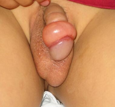
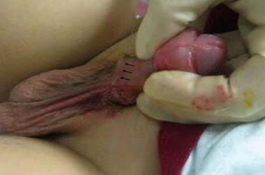
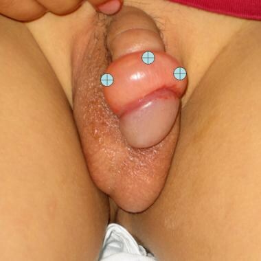
Paraphimosis is the inability to reduce a swollen and proximally positioned foreskin over the glans penis (see images below). [1, 2, 3, 4] Paraphimosis is most often iatrogenic, occurring when medical personnel forget to reduce the foreskin after instrumentation or catheterization of the urethra. [1, 5] The foreskin does not become fully mobile before the age of 3-4 years, predisposing children younger than 3-4 years to paraphimosis when their caregivers retract the foreskin for cleaning. Cases of paraphimosis as a complication of monkeypox (mpox) have also been reported. [6, 7]
Patients present with a red, painful, and swollen glans penis associated with an edematous, proximally retracted foreskin that forms a circumferential constricting band. The penile shaft proximal to the constricting band typically is soft. [8]
The retracted foreskin initially blocks lymphatic drainage from the distal penis, progressively causing further edema of the retracted foreskin. If the foreskin remains retracted and the edema continuous, venous obstruction followed by arterial flow are expected within hours to days. [9]
All patients with paraphimosis require emergent reduction.
For more information on paraphimosis and the related condition phimosis, see Medscape Reference article Phimosis and Paraphimosis.
Contraindications
Nonsurgical techniques are relatively contraindicated in patients who have the following conditions:
-
Necrotic or ulcerated foreskin
-
Necrotic or ulcerated penis
Anesthesia
Reduction of paraphimosis is a painful procedure. The use of parenteral analgesic and sedative agents is recommended. Anesthesia with local infiltration may be used.
Various methods of penile anesthesia exist (see Technique section). For more information, see Nerve Block, Dorsal Penile.
Procedural sedation is recommended in the pediatric patient. For more information, see Procedural Sedation.
Equipment
Equipment may include the following:
-
Topical anesthetic cream (eutectic mixture of local anesthetics [EMLA]; lidocaine, adrenaline, tetracaine [LAT]; tetracaine, adrenaline, cocaine [TAC]) (For more information, see Anesthesia, Topical.) [10]
-
4 x 4 gauze
-
Povidone-iodine solution (eg, Betadine)
-
Sterile drapes
-
Sterile gloves
-
Local anesthetic solution without epinephrine (lidocaine 1%)
-
Syringe, 10 mL
-
Needles, 18- and 27-gauge (ga)
-
Crushed ice
-
Babcock clamps, 6-8
Positioning and Technique
Positioning
Position the patient supine, with his legs separated.
Preparation and anesthesia
Preparation and anesthesia include the following steps:
-
Obtain informed consent. Explain the procedure in detail to the parent/guardian of a pediatric patient.
-
Apply a liberal amount of the local anesthetic cream to the glans and foreskin.
-
Wait for the anesthesia to take effect.
-
Clean the penis of the local anesthetic cream.
-
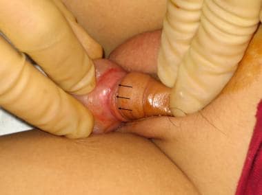
Apply an antiseptic solution to the penis and foreskin (see image below).
 Application of antiseptic solution and identification of the superficial dorsal penile vein.
-
Place sterile drapes to create a sterile field around the penis.
-
If adequate anesthesia has not been achieved, penile anesthesia should be performed. One of two techniques may be used for penile anesthesia. Both techniques are painful and require the administration of parenteral analgesics and/or procedural sedation and analgesia.
Inject 1-2 mL of lidocaine 1% near the right and left dorsal penile nerves (the 10- and 2-o'clock positions close to the base of the penis; see image below).
Circumferentially infiltrate lidocaine 1% around the base of the penis (see image below).

Manual reduction
Steps in manual reduction include the following:
-
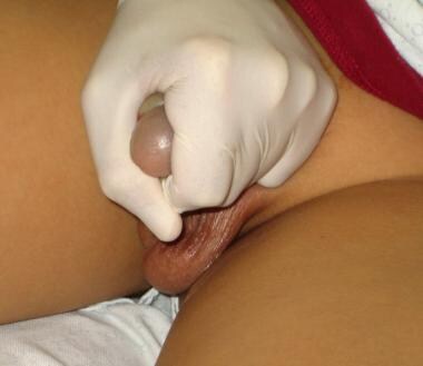
Apply slow and steady manual compression over the glans penis and edematous foreskin, squeezing distally to proximally in order to mobilize the edema proximally (see image below).
-
The pressure should be applied for 5-10 minutes and can be delegated to the patient or caregiver.
-
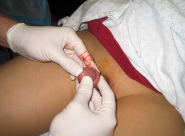
After manual compression, position the thumbs on both sides of the urethral meatus and the index and middle fingers proximal to the phimotic ring (see image below).
-
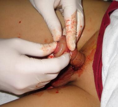
Apply continuous force to move the phimotic ring distally over the glans (see image below).
Iced glove technique
The steps involved in the technique are as follows:
-
Half fill a surgical glove with water and ice chips, express the remaining air, and tie a knot in the wrist of the glove.
-
Insert the penis into the invaginated thumb of the glove.
-
Apply circumferential pressure for 5-10 minutes.
-
Attempt to manually reduce the foreskin again.
Babcock clamp technique
The following steps are involved in the technique:
-
This technique requires administration of local anesthetic.
-
The Babcock clamp is a noncrushing tissue clamp.
It can be safely used to reduce the paraphimotic foreskin.
All other clamps will crush the foreskin tissue.
-
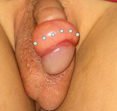
Apply 6-8 Babcock clamps evenly spaced around the foreskin by placing one edge just proximal to the phimotic ring with the other edge just distal to the phimotic ring (see image below).
-
Grasp all clamps in one hand, and simultaneously apply distal traction to pull the phimotic ring over the glans.
-
After reduction, remove the clamps and inspect the foreskin for injuries.
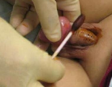
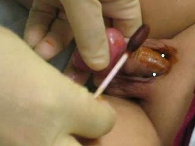
Needle decompression technique
The steps involved in the technique are as follows:
-
This technique requires administration of local anesthetic.
-
Clean the penis, apply antiseptic solution, and place drapes to create a sterile field.
-
Use an 18-gauge needle to create 8-12 circumferential holes, 3-5 mm deep, in the foreskin (see image below).
-
Wrap a piece of 4 x 4 gauze around the glans and foreskin, and apply manual compression for 5-10 minutes, allowing the foreskin to decompress by draining edematous fluid and blood.
-
Attempt to manually reduce the foreskin again.
Aftercare
Aftercare includes the following:
-
The successfully reduced foreskin should look like a normal uncircumcised penis (see image below).
 The successfully reduced paraphimosis should have the appearance of a normal uncircumcised penis.
-
The patient should feel relief of pressure and pain.
-
Residual swelling is expected to resolve in a few days.
-
Observe the patient to ensure the following:
Recovery from anesthesia
Adequate hemostasis
Ability to urinate
-
All patients should be referred to a urologist in 1-2 days.

Pearls
Apply slow and steady manual compression over the glans penis and edematous foreskin, squeezing distally to proximally in order to mobilize the edema proximally. This pressure should be applied for 5-10 minutes and can be delegated to the patient or caregiver.
Circumcision is the recommended definitive therapy for most patients who experience non-iatrogenic paraphimosis. [11, 12] For more information on circumcision in children and adults, see Medscape Reference Pediatrics article Circumcision and Urology article Phimosis, Adult Circumcision, and Buried Penis.
Complications
Complications may include the following:
-
A significant degree of pain after the procedure is uncommon. If it occurs, it can be treated with a topical anesthetic.
-
Swelling usually takes a few days to resolve.
-
A dorsal slit of the foreskin is indicated if less invasive techniques fail to achieve reduction. [2, 13]
-
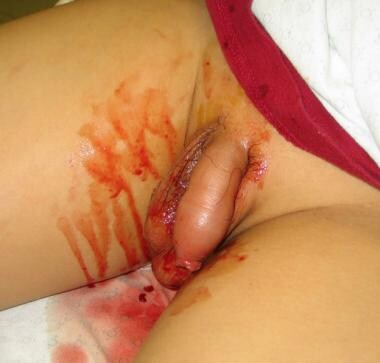
Penile or foreskin lacerations or tears that result from the manual reduction should be sutured with an absorbable material.
-
Some bleeding is common following needle decompression and dorsal slit techniques. A compressive dressing may aid with hemostasis.
-
Prescribing antibiotics to patients with evidence of skin infection or ulcers or after any invasive procedure that involves the foreskin is recommended.
-
Paraphimosis.
-
Application of antiseptic solution and identification of the superficial dorsal penile vein.
-
Anesthetic injection sites in the 10- and 2-o'clock positions.
-
Circumferential infiltration of local anesthetic.
-
Manual compression of the glans and foreskin.
-
Manual reduction of paraphimosis.
-
Manual reduction of paraphimosis.
-
Constricting (phimotic) ring.
-
The successfully reduced paraphimosis should have the appearance of a normal uncircumcised penis.
-
The Babcock clamps should be placed along the phimotic ring.
-
An 18-gauge needle is used to circumferentially puncture the edematous foreskin.
-
Manual reduction of paraphimosis.


