Having caused an extensive health scare and over 1,000 deaths so far, the COVID-19 virus (also unofficially known as 2019-nCoV) has received wide media coverage since its discovery in December last year.
But although microbiologists around the world have been using the virus to try and develop a vaccine, many of us non-scientists haven't actually seen what this new coronavirus looks like.
The National Institute of Allergy and Infectious Diseases' (NIAID) Rocky Mountains Laboratories (RML) have just released scanning and transmission electron microscope images of the coronavirus, and they're… surprisingly aesthetically pleasing.
 (NIAID-RML)
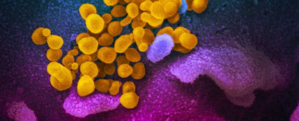
What you're seeing above is a scanning electron microscope image in false colour, showing the COVID-19 virus from a patient in the US; the viral particles are coloured yellow as it emerges from the surface of a cell, which is coloured blue and pink.
 (NIAID-RML)
The image above was captured with a transmission electron microscope. This isn't quite as sharp as the first one, but you can see the spikes on the surface of the virus (which gives the coronavirus its name, meaning 'crown').
If these look a little familiar, it's because most coronaviruses – such as SARS and MERS - look similar on the outside, sharing the bump-covered spherical appearance.
Viruses in the coronavirus family only have small differences in their genome, with only five nucleotide differences between three of the viruses. Nevertheless, the viruses can have notably different presentations when it comes to infecting humans.
 (NIAID-RML)
The images you see here were the result of collaborative teamwork. RML investigator Emmie de Wit provided the virus, microscopist Elizabeth Fischer produced the images, with the visual medical arts office colourising the images.
You can see the entire set on Flickr here.